 Movie
Movie Controller

[English] 日本語
 Yorodumi
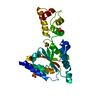
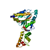
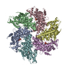
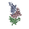
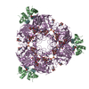
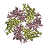
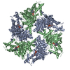
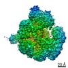
- PDB-2dhr: Whole cytosolic region of ATP-dependent metalloprotease FtsH (G399L) -
+ Open data

- Basic information
| Entry | Database: PDB / ID: 2dhr | ||||||
|---|---|---|---|---|---|---|---|
| Title | Whole cytosolic region of ATP-dependent metalloprotease FtsH (G399L) | ||||||
 Components Components | FtsH | ||||||
 Keywords Keywords | HYDROLASE / AAA+ protein / hexameric Zn metalloprotease | ||||||
| Function / homology |  Function and homology information Function and homology informationHydrolases; Acting on peptide bonds (peptidases); Metalloendopeptidases / ATP-dependent peptidase activity / protein catabolic process / metalloendopeptidase activity / ATP hydrolysis activity / proteolysis / zinc ion binding / ATP binding / plasma membrane Similarity search - Function | ||||||
| Biological species |   Thermus thermophilus (bacteria) Thermus thermophilus (bacteria) | ||||||
| Method |  X-RAY DIFFRACTION / SYNCHROTRON / MIR / Resolution: 3.9 Å X-RAY DIFFRACTION / SYNCHROTRON / MIR / Resolution: 3.9 Å | ||||||
 Authors Authors | Suno, R. / Niwa, H. / Tsuchiya, D. / Zhang, X. / Yoshida, M. / Morikawa, K. | ||||||
 Citation Citation | Journal: Mol.Cell / Year: 2006 Title: Structure of the Whole Cytosolic Region of ATP-Dependent Protease FtsH Authors: Suno, R. / Niwa, H. / Tsuchiya, D. / Zhang, X. / Yoshida, M. / Morikawa, K. #1: Journal: Structure / Year: 2002Title: Hexameric ring structure of the ATPase domain of the membrane-integrated metalloprotease FtsH from Thermus thermophilus HB8 Authors: Niwa, H. / Tsuchiya, D. / Makyio, H. / Yoshida, M. / Morikawa, K. | ||||||
| History |
|
- Structure visualization
| Structure viewer | Molecule: MolmilJmol/JSmol |
|---|
- Downloads & links
-Download
| PDBx/mmCIF format | 2dhr.cif.gz | 521.7 KB | Display | PDBx/mmCIF format |
|---|---|---|---|---|
| PDB format | pdb2dhr.ent.gz | 425.4 KB | Display | PDB format |
| PDBx/mmJSON format | 2dhr.json.gz | Tree view | PDBx/mmJSON format | |
| Others |  Other downloads Other downloads |
-Validation report
| Arichive directory | https://data.pdbj.org/pub/pdb/validation_reports/dh/2dhrftp://data.pdbj.org/pub/pdb/validation_reports/dh/2dhr | HTTPS FTP |
|---|
-Related structure data


-Links
 PDBj
PDBj


- Assembly
| Deposited unit | 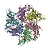
| ||||||||
|---|---|---|---|---|---|---|---|---|---|
| 1 |
| ||||||||
| Unit cell |
| ||||||||
| Details | The biological assembly is a hexamer composed of the six polypeptide chains in the asymmetric unit. |
-Components
| #1: Protein | Mass: 55369.281 Da / Num. of mol.: 6 / Fragment: whole cytosolic region / Mutation: G399L Source method: isolated from a genetically manipulated source Source: (gene. exp.) Thermus thermophilus (bacteria) / Production host: #2: Chemical | ChemComp-ADP /   Mass: 427.201 Da / Num. of mol.: 6 / Source method: obtained synthetically / Formula: C10H15N5O10P2 / Comment: ADP, energy-carrying molecule*YM Mass: 427.201 Da / Num. of mol.: 6 / Source method: obtained synthetically / Formula: C10H15N5O10P2 / Comment: ADP, energy-carrying molecule*YM |
|---|
-Experimental details
-Experiment
| Experiment | Method: X-RAY DIFFRACTION / Number of used crystals: 1 |
|---|
- Sample preparation
| Crystal | Density Matthews: 3.24 Å3/Da / Density % sol: 62.01 % |
|---|---|
| Crystal grow | Temperature: 293 K / Method: vapor diffusion, hanging drop / pH: 7.2 Details: 0.1M Tris-HCl, 1% PEG8K, 0.2mM Zn acetate, 2.5mM ADP, pH 7.2, VAPOR DIFFUSION, HANGING DROP, temperature 293K |
-Data collection
| Diffraction | Mean temperature: 100 K |
|---|---|
| Diffraction source | Source: SYNCHROTRON / Site: SPring-8  / Beamline: BL41XU / Wavelength: 1 Å / Beamline: BL41XU / Wavelength: 1 Å |
| Detector | Type: ADSC QUANTUM 315 / Detector: CCD / Date: May 20, 2005 |
| Radiation | Monochromator: Si / Protocol: SINGLE WAVELENGTH / Monochromatic (M) / Laue (L): M / Scattering type: x-ray |
| Radiation wavelength | Wavelength: 1 Å / Relative weight: 1 |
| Reflection | Resolution: 3.9→100 Å / Num. all: 40304 / Num. obs: 40304 / % possible obs: 99.9 % / Observed criterion σ(F): 0 / Observed criterion σ(I): 0 / Rmerge(I) obs: 0.077 |
| Reflection shell | Resolution: 3.9→4.1 Å / Rmerge(I) obs: 0.547 / % possible all: 100 |
- Processing
| Software |
| ||||||||||||||||||||
|---|---|---|---|---|---|---|---|---|---|---|---|---|---|---|---|---|---|---|---|---|---|
| Refinement | Method to determine structure: MIR / Resolution: 3.9→15 Å / Cross valid method: THROUGHOUT / σ(F): 3 / Stereochemistry target values: Engh & Huber
| ||||||||||||||||||||
| Refinement step | Cycle: LAST / Resolution: 3.9→15 Å
|
