 Movie
Movie Controller

[English] 日本語
 Yorodumi
- PDB-2d1z: Crystal structure of catalytic-site mutant xylanase from Streptom... -
+ Open data

- Basic information
| Entry | Database: PDB / ID: 2d1z | ||||||
|---|---|---|---|---|---|---|---|
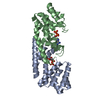
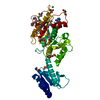
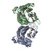
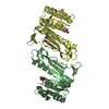
| Title | Crystal structure of catalytic-site mutant xylanase from Streptomyces olivaceoviridis E-86 | ||||||
 Components Components | ENDO-1,4-BETA-D-XYLANASE | ||||||
 Keywords Keywords | HYDROLASE / TIM-BARREL / RETAINING ENZYME / CATALYTIC-SITE MUTANT / CHEMICAL RESCUE | ||||||
| Function / homology |  Function and homology information Function and homology informationendo-1,4-beta-xylanase / endo-1,4-beta-xylanase activity / xylan catabolic process Similarity search - Function | ||||||
| Biological species |  Streptomyces olivaceoviridis (bacteria) Streptomyces olivaceoviridis (bacteria) | ||||||
| Method |  X-RAY DIFFRACTION / SYNCHROTRON / MOLECULAR REPLACEMENT / Resolution: 1.6 Å X-RAY DIFFRACTION / SYNCHROTRON / MOLECULAR REPLACEMENT / Resolution: 1.6 Å | ||||||
 Authors Authors | Suzuki, R. / Kuno, A. / Fujimoto, Z. / Ito, S. / Kawahara, S.I. / Kaneko, S. / Hasegawa, T. / Taira, K. | ||||||
 Citation Citation | Journal: J.Biochem. / Year: 2009 Title: Crystallographic snapshots of an entire reaction cycle for a retaining xylanase from Streptomyces olivaceoviridis E-86 Authors: Suzuki, R. / Fujimoto, Z. / Ito, S. / Kawahara, S. / Kaneko, S. / Taira, K. / Hasegawa, T. / Kuno, A. #1: Journal: J.Mol.Biol. / Year: 2000Title: Crystal structure of Streptomyces olivaceoviridis E-86 beta-xylanase containing xylan-binding domain Authors: Fujimoto, Z. / Kuno, A. / Kaneko, S. / Yoshida, S. / Kobayashi, H. / Kusakabe, I. / Mizuno, H. #2: Journal: J.Mol.Biol. / Year: 2002Title: Crystal structures of the sugar complexes of Streptomyces olivaceoviridis E-86 xylanase: sugar binding structure of the family 13 carbohydrate binding module Authors: Fujimoto, Z. / Kuno, A. / Kaneko, S. / Kobayashi, H. / Kusakabe, I. / Mizuno, H. #3: Journal: J.Biol.Chem. / Year: 2004Title: Crystal structures of decorated xylooligosaccharides bound to a family 10 xylanase from Streptomyces olivaceoviridis E-86 Authors: Fujimoto, Z. / Kaneko, S. / Kuno, A. / Kobayashi, H. / Kusakabe, I. / Mizuno, H. #4: Journal: J.FERMENT.BIOENG. / Year: 1998Title: PCR Cloning and Expression of the F/10 Family Xylanse Gene from Streptomyces olivaceoviridis E-86 Authors: Kuno, A. / Shimizu, D. / Kaneko, S. / Koyama, Y. / Yoshida, S. / Kobayashi, H. / Hayashi, K. / Taira, K. / Kusakabe, I. #5: Journal: Febs Lett. / Year: 1999 Title: Significant enhancement in the binding of p-nitrophenyl-beta-D-xylobioside by the E128H mutant F/10 xylanase from Streptomyces olivaceoviridis E-86 Authors: Kuno, A. / Shimizu, D. / Kaneko, S. / Hasegawa, T. / Gama, Y. / Hayashi, K. / Kusakabe, I. / Taira, K. | ||||||
| History |
|
- Structure visualization
| Structure viewer | Molecule: MolmilJmol/JSmol |
|---|
- Downloads & links
-Download
| PDBx/mmCIF format | 2d1z.cif.gz | 195.9 KB | Display | PDBx/mmCIF format |
|---|---|---|---|---|
| PDB format | pdb2d1z.ent.gz | 152.5 KB | Display | PDB format |
| PDBx/mmJSON format | 2d1z.json.gz | Tree view | PDBx/mmJSON format | |
| Others |  Other downloads Other downloads |
-Validation report
| Arichive directory | https://data.pdbj.org/pub/pdb/validation_reports/d1/2d1zftp://data.pdbj.org/pub/pdb/validation_reports/d1/2d1z | HTTPS FTP |
|---|
-Related structure data
| Related structure data |  2d20C  2d22C  2d23C  2d24C  1xyfS S: Starting model for refinement C: citing same article ( |
|---|---|
| Similar structure data |


-Links
 PDBj
PDBj


- Assembly
| Deposited unit | 
| ||||||||
|---|---|---|---|---|---|---|---|---|---|
| 1 | 
| ||||||||
| 2 | 
| ||||||||
| Unit cell |
|
-Components
| #1: Protein | Mass: 46773.230 Da / Num. of mol.: 2 / Mutation: N127S and E128H Source method: isolated from a genetically manipulated source Source: (gene. exp.) Streptomyces olivaceoviridis (bacteria)Strain: E-86 / Plasmid: pQE60 / Production host: #2: Chemical | ChemComp-SO4 / |   Mass: 96.063 Da / Num. of mol.: 1 / Source method: obtained synthetically / Formula: SO4 Mass: 96.063 Da / Num. of mol.: 1 / Source method: obtained synthetically / Formula: SO4#3: Chemical | ChemComp-GOL /   Mass: 92.094 Da / Num. of mol.: 11 / Source method: obtained synthetically / Formula: C3H8O3 Mass: 92.094 Da / Num. of mol.: 11 / Source method: obtained synthetically / Formula: C3H8O3#4: Water | ChemComp-HOH / |  Mass: 18.015 Da / Num. of mol.: 901 / Source method: isolated from a natural source / Formula: H2O Mass: 18.015 Da / Num. of mol.: 901 / Source method: isolated from a natural source / Formula: H2OHas protein modification | Y | |
|---|
-Experimental details
-Experiment
| Experiment | Method: X-RAY DIFFRACTION / Number of used crystals: 1 |
|---|
- Sample preparation
| Crystal | Density Matthews: 2.9 Å3/Da / Density % sol: 56.7 % |
|---|---|
| Crystal grow | Temperature: 293 K / Method: vapor diffusion, hanging drop / pH: 6.5 Details: disodium hydrogenphosphate, citric acid, ammonium sulfate, pH 6.5, VAPOR DIFFUSION, HANGING DROP, temperature 293K |
-Data collection
| Diffraction | Mean temperature: 95 K |
|---|---|
| Diffraction source | Source: SYNCHROTRON / Site: Photon Factory  / Beamline: AR-NW12A / Wavelength: 0.978 Å / Beamline: AR-NW12A / Wavelength: 0.978 Å |
| Detector | Type: ADSC QUANTUM 210 / Detector: CCD / Date: Dec 3, 2003 |
| Radiation | Monochromator: Si(111) double crystal monochromator / Protocol: SINGLE WAVELENGTH / Monochromatic (M) / Laue (L): M / Scattering type: x-ray |
| Radiation wavelength | Wavelength: 0.978 Å / Relative weight: 1 |
| Reflection | Resolution: 1.6→50 Å / Num. all: 260493 / Num. obs: 136080 / % possible obs: 99.4 % / Observed criterion σ(I): 0 / Biso Wilson estimate: 18.2 Å2 |
| Reflection shell | Resolution: 1.6→1.66 Å / % possible all: 97.1 |
- Processing
| Software |
| |||||||||||||||||||||||||
|---|---|---|---|---|---|---|---|---|---|---|---|---|---|---|---|---|---|---|---|---|---|---|---|---|---|---|
| Refinement | Method to determine structure: MOLECULAR REPLACEMENT Starting model: PDB ENTRY 1XYF Resolution: 1.6→47.03 Å / Rfactor Rfree error: 0.002 / Data cutoff high absF: 2478062.82 / Data cutoff low absF: 0 / Isotropic thermal model: RESTRAINED / Cross valid method: THROUGHOUT / σ(F): 0 / Stereochemistry target values: Engh & Huber
| |||||||||||||||||||||||||
| Solvent computation | Solvent model: FLAT MODEL / Bsol: 47.4026 Å2 / ksol: 0.390252 e/Å3 | |||||||||||||||||||||||||
| Displacement parameters | Biso mean: 19.6 Å2
| |||||||||||||||||||||||||
| Refine analyze |
| |||||||||||||||||||||||||
| Refinement step | Cycle: LAST / Resolution: 1.6→47.03 Å
| |||||||||||||||||||||||||
| Refine LS restraints |
| |||||||||||||||||||||||||
| LS refinement shell | Resolution: 1.6→1.7 Å / Rfactor Rfree error: 0.007 / Total num. of bins used: 6
| |||||||||||||||||||||||||
| Xplor file |
|
