 Movie
Movie Controller

[English] 日本語
 Yorodumi
- PDB-1isw: Crystal structure of xylanase from Streptomyces olivaceoviridis E... -
+ Open data

- Basic information
| Entry | Database: PDB / ID: 1isw | |||||||||
|---|---|---|---|---|---|---|---|---|---|---|
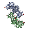
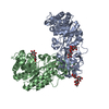
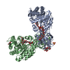
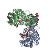
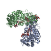
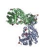
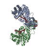
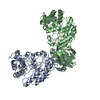
| Title | Crystal structure of xylanase from Streptomyces olivaceoviridis E-86 complexed with xylobiose | |||||||||
 Components Components | endo-1,4-beta-D-xylanase | |||||||||
 Keywords Keywords | HYDROLASE / ALPHA-BETA BARREL / PROTEIN-SUGAR COMPLEX / CARBOHYDRATE BINDING DOMAIN | |||||||||
| Function / homology |  Function and homology information Function and homology informationendo-1,4-beta-xylanase / endo-1,4-beta-xylanase activity / xylan catabolic process Similarity search - Function | |||||||||
| Biological species |  Streptomyces olivaceoviridis (bacteria) Streptomyces olivaceoviridis (bacteria) | |||||||||
| Method |  X-RAY DIFFRACTION / MOLECULAR REPLACEMENT / Resolution: 2.1 Å X-RAY DIFFRACTION / MOLECULAR REPLACEMENT / Resolution: 2.1 Å | |||||||||
 Authors Authors | Fujimoto, Z. / Kuno, A. / Kaneko, S. / Kobayashi, H. / Kusakabe, I. / Mizuno, H. | |||||||||
 Citation Citation | Journal: J.Mol.Biol. / Year: 2002 Title: Crystal structures of the sugar complexes of Streptomyces olivaceoviridis E-86 xylanase: sugar binding structure of the family 13 carbohydrate binding module. Authors: Fujimoto, Z. / Kuno, A. / Kaneko, S. / Kobayashi, H. / Kusakabe, I. / Mizuno, H. #1: Journal: J.Mol.Biol. / Year: 2000Title: Crystal structure of Streptomyces olivaceoviridis E-86 beta-xylanase containing xylan-binding domain Authors: Fujimoto, Z. / Kuno, A. / Kaneko, S. / Yoshida, S. / Kobayashi, H. / Kusakabe, I. / Mizuno, H. #2: Journal: J.BIOCHEM.(TOKYO) / Year: 1997Title: Crystallization and preliminary X-ray crystallographic study of Streptomyces olivaceoviridis E-86 beta-xylanase Authors: Fujimoto, Z. / Mizuno, H. / Kuno, A. / Yoshida, S. / Kobayashi, H. / Kusakabe, I. | |||||||||
| History |
|
- Structure visualization
| Structure viewer | Molecule: MolmilJmol/JSmol |
|---|
- Downloads & links
-Download
| PDBx/mmCIF format | 1isw.cif.gz | 195 KB | Display | PDBx/mmCIF format |
|---|---|---|---|---|
| PDB format | pdb1isw.ent.gz | 153.4 KB | Display | PDB format |
| PDBx/mmJSON format | 1isw.json.gz | Tree view | PDBx/mmJSON format | |
| Others |  Other downloads Other downloads |
-Validation report
| Arichive directory | https://data.pdbj.org/pub/pdb/validation_reports/is/1iswftp://data.pdbj.org/pub/pdb/validation_reports/is/1isw | HTTPS FTP |
|---|
-Related structure data
| Related structure data | 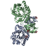 1isvC  1isxC 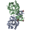 1isyC  1iszC 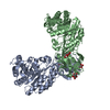 1it0C  1xyfS S: Starting model for refinement C: citing same article ( |
|---|---|
| Similar structure data |


-Links
 PDBj
PDBj


- Assembly
| Deposited unit | 
| ||||||||
|---|---|---|---|---|---|---|---|---|---|
| 1 |
| ||||||||
| Unit cell |
|
-Components
| #1: Protein | Mass: 46791.223 Da / Num. of mol.: 2 Source method: isolated from a genetically manipulated source Source: (gene. exp.) Streptomyces olivaceoviridis (bacteria)Strain: E-86 / Production host: #2: Polysaccharide | beta-D-xylopyranose-(1-4)-beta-D-xylopyranose / 4beta-beta-xylobiose 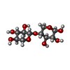  Source method: isolated from a genetically manipulated source Details: oligosaccharide / References: 4beta-beta-xylobiose #3: Sugar | ChemComp-XYP /   Type: D-saccharide, beta linking / Mass: 150.130 Da / Num. of mol.: 4 Type: D-saccharide, beta linking / Mass: 150.130 Da / Num. of mol.: 4Source method: isolated from a genetically manipulated source Formula: C5H10O5 #4: Water | ChemComp-HOH / |  Mass: 18.015 Da / Num. of mol.: 740 / Source method: isolated from a natural source / Formula: H2O Mass: 18.015 Da / Num. of mol.: 740 / Source method: isolated from a natural source / Formula: H2OHas protein modification | Y | |
|---|
-Experimental details
-Experiment
| Experiment | Method: X-RAY DIFFRACTION / Number of used crystals: 1 |
|---|
- Sample preparation
| Crystal | Density Matthews: 2.6 Å3/Da / Density % sol: 52.77 % | ||||||||||||||||||||||||||||||
|---|---|---|---|---|---|---|---|---|---|---|---|---|---|---|---|---|---|---|---|---|---|---|---|---|---|---|---|---|---|---|---|
| Crystal grow | Temperature: 298 K / Method: vapor diffusion, hanging drop / pH: 5.7 Details: ammonium sulfate, pH 5.7, VAPOR DIFFUSION, HANGING DROP, temperature 298K | ||||||||||||||||||||||||||||||
| Crystal grow | *PLUS | ||||||||||||||||||||||||||||||
| Components of the solutions | *PLUS
|
-Data collection
| Diffraction | Mean temperature: 100 K |
|---|---|
| Diffraction source | Source: ROTATING ANODE / Type: RIGAKU RU300 / Wavelength: 1.5418 Å |
| Detector | Type: RIGAKU RAXIS IV / Detector: IMAGE PLATE / Date: May 12, 2001 |
| Radiation | Protocol: SINGLE WAVELENGTH / Monochromatic (M) / Laue (L): M / Scattering type: x-ray |
| Radiation wavelength | Wavelength: 1.5418 Å / Relative weight: 1 |
| Reflection | Resolution: 2→46 Å / Num. all: 359683 / Num. obs: 66842 / % possible obs: 86.6 % / Observed criterion σ(F): 2 / Observed criterion σ(I): 2 / Redundancy: 5.9 % / Biso Wilson estimate: 19.4 Å2 / Rmerge(I) obs: 0.067 / Net I/σ(I): 19.4 |
| Reflection shell | Resolution: 2→2.07 Å / Redundancy: 6.22 % / Rmerge(I) obs: 0.316 / Mean I/σ(I) obs: 6.4 / Num. unique all: 6586 / % possible all: 77.7 |
| Reflection | *PLUS Num. measured all: 359683 |
- Processing
| Software |
| ||||||||||||||||||||||||||||||||||||||||||||||||||||||||||||||||||||||||||||||||
|---|---|---|---|---|---|---|---|---|---|---|---|---|---|---|---|---|---|---|---|---|---|---|---|---|---|---|---|---|---|---|---|---|---|---|---|---|---|---|---|---|---|---|---|---|---|---|---|---|---|---|---|---|---|---|---|---|---|---|---|---|---|---|---|---|---|---|---|---|---|---|---|---|---|---|---|---|---|---|---|---|---|
| Refinement | Method to determine structure: MOLECULAR REPLACEMENT Starting model: PDB ENTRY 1XYF Resolution: 2.1→29.74 Å / Rfactor Rfree error: 0.003 / Isotropic thermal model: RESTRAINED / Cross valid method: THROUGHOUT / σ(F): 0
| ||||||||||||||||||||||||||||||||||||||||||||||||||||||||||||||||||||||||||||||||
| Solvent computation | Solvent model: FLAT MODEL / Bsol: 66.7471 Å2 / ksol: 0.39314 e/Å3 | ||||||||||||||||||||||||||||||||||||||||||||||||||||||||||||||||||||||||||||||||
| Displacement parameters |
| ||||||||||||||||||||||||||||||||||||||||||||||||||||||||||||||||||||||||||||||||
| Refine analyze |
| ||||||||||||||||||||||||||||||||||||||||||||||||||||||||||||||||||||||||||||||||
| Refinement step | Cycle: LAST / Resolution: 2.1→29.74 Å
| ||||||||||||||||||||||||||||||||||||||||||||||||||||||||||||||||||||||||||||||||
| Refine LS restraints |
| ||||||||||||||||||||||||||||||||||||||||||||||||||||||||||||||||||||||||||||||||
| LS refinement shell | Resolution: 2.1→2.23 Å / Rfactor Rfree error: 0.01 / Total num. of bins used: 6
| ||||||||||||||||||||||||||||||||||||||||||||||||||||||||||||||||||||||||||||||||
| Xplor file |
| ||||||||||||||||||||||||||||||||||||||||||||||||||||||||||||||||||||||||||||||||
| Software | *PLUS Name: CNS / Version: 1 / Classification: refinement | ||||||||||||||||||||||||||||||||||||||||||||||||||||||||||||||||||||||||||||||||
| Refinement | *PLUS Highest resolution: 2.1 Å / Lowest resolution: 30 Å / σ(F): 0 / % reflection Rfree: 10.2 % / Rfactor obs: 0.196 / Rfactor Rfree: 0.244 | ||||||||||||||||||||||||||||||||||||||||||||||||||||||||||||||||||||||||||||||||
| Solvent computation | *PLUS | ||||||||||||||||||||||||||||||||||||||||||||||||||||||||||||||||||||||||||||||||
| Displacement parameters | *PLUS Biso mean: 28.2 Å2 | ||||||||||||||||||||||||||||||||||||||||||||||||||||||||||||||||||||||||||||||||
| Refine LS restraints | *PLUS
| ||||||||||||||||||||||||||||||||||||||||||||||||||||||||||||||||||||||||||||||||
| LS refinement shell | *PLUS Highest resolution: 2.1 Å / Lowest resolution: 2.18 Å / % reflection Rfree: 11.1 % / Rfactor Rwork: 0.244 / Num. reflection Rwork: 4447 / Rfactor obs: 0.244 |
