 Movie
Movie Controller

[English] 日本語
 Yorodumi
- PDB-5gqe: Crystal structure of michaelis complex of xylanase mutant (T82A, ... -
+ Open data

- Basic information
| Entry | Database: PDB / ID: 5gqe | |||||||||
|---|---|---|---|---|---|---|---|---|---|---|
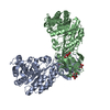
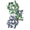
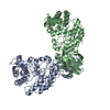
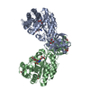
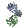
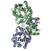
| Title | Crystal structure of michaelis complex of xylanase mutant (T82A, N127S, and E128H) from Streptomyces olivaceoviridis E-86 | |||||||||
 Components Components | Beta-xylanase | |||||||||
 Keywords Keywords | HYDROLASE / XYLOPENTAOSE / ES COMPLEX | |||||||||
| Function / homology |  Function and homology information Function and homology informationendo-1,4-beta-xylanase / endo-1,4-beta-xylanase activity / xylan catabolic process Similarity search - Function | |||||||||
| Biological species |  Streptomyces olivaceoviridis (bacteria) Streptomyces olivaceoviridis (bacteria) | |||||||||
| Method |  X-RAY DIFFRACTION / MOLECULAR REPLACEMENT / Resolution: 2.5 Å X-RAY DIFFRACTION / MOLECULAR REPLACEMENT / Resolution: 2.5 Å | |||||||||
 Authors Authors | Suzuki, R. / Fujimoto, Z. / Kaneko, S. / Kuno, A. | |||||||||
 Citation Citation | Journal: J.Appl.Glyosci. / Year: 2019 Title: Azidolysis by the Formation of Stable Ser-His Catalytic Dyad in a Glycoside Hydrolase Family 10 Xylanase Mutant Authors: Suzuki, R. / Fujimoto, Z. / Kaneko, S. / Hasegawa, T. / Kuno, A. #1: Journal: J. Biochem. / Year: 2009Title: Crystallographic snapshots of an entire reaction cycle for a retaining xylanase from Streptomyces olivaceoviridis E-86. Authors: Suzuki, R. / Fujimoto, Z. / Ito, S. / Kawahara, S. / Kaneko, S. / Taira, K. / Hasegawa, T. / Kuno, A. | |||||||||
| History |
|
- Structure visualization
| Structure viewer | Molecule: MolmilJmol/JSmol |
|---|
- Downloads & links
-Download
| PDBx/mmCIF format | 5gqe.cif.gz | 196.4 KB | Display | PDBx/mmCIF format |
|---|---|---|---|---|
| PDB format | pdb5gqe.ent.gz | 153.2 KB | Display | PDB format |
| PDBx/mmJSON format | 5gqe.json.gz | Tree view | PDBx/mmJSON format | |
| Others |  Other downloads Other downloads |
-Validation report
| Arichive directory | https://data.pdbj.org/pub/pdb/validation_reports/gq/5gqeftp://data.pdbj.org/pub/pdb/validation_reports/gq/5gqe | HTTPS FTP |
|---|
-Related structure data
| Related structure data |  5gqdC  2d1zS C: citing same article ( S: Starting model for refinement |
|---|---|
| Similar structure data |

-Links
 PDBj
PDBj


- Assembly
| Deposited unit | 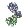
| ||||||||
|---|---|---|---|---|---|---|---|---|---|
| 1 |
| ||||||||
| 2 | 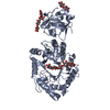
| ||||||||
| 3 | 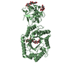
| ||||||||
| Unit cell |
|
-Components
-Protein / Non-polymers , 2 types, 729 molecules AB
| #1: Protein | Mass: 46743.207 Da / Num. of mol.: 2 Source method: isolated from a genetically manipulated source Source: (gene. exp.) Streptomyces olivaceoviridis (bacteria)Production host: #6: Water | ChemComp-HOH / | Mass: 18.015 Da / Num. of mol.: 727 / Source method: isolated from a natural source / Formula: H2O |
|---|
-Sugars , 4 types, 6 molecules
| #2: Polysaccharide | Source method: isolated from a genetically manipulated source #3: Polysaccharide | beta-D-xylopyranose-(1-4)-beta-D-xylopyranose-(1-4)-beta-D-xylopyranose-(1-4)-beta-D-xylopyranose | Source method: isolated from a genetically manipulated source #4: Polysaccharide | 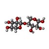  Source method: isolated from a genetically manipulated source Details: oligosaccharide / References: 4beta-beta-xylobiose #5: Polysaccharide | beta-D-xylopyranose-(1-4)-beta-D-xylopyranose-(1-4)-beta-D-xylopyranose / 4beta-beta-xylotriose |   Source method: isolated from a genetically manipulated source Details: oligosaccharide / References: 4beta-beta-xylotriose |
|---|
-Details
| Has protein modification | Y |
|---|
-Experimental details
-Experiment
| Experiment | Method: X-RAY DIFFRACTION / Number of used crystals: 1 |
|---|
- Sample preparation
| Crystal | Density Matthews: 2.7 Å3/Da / Density % sol: 54.44 % |
|---|---|
| Crystal grow | Temperature: 293 K / Method: vapor diffusion, hanging drop / pH: 5.7 / Details: 2% McIlvaine buffer 25% Ammonium sulfate |
-Data collection
| Diffraction | Mean temperature: 173 K |
|---|---|
| Diffraction source | Source: ROTATING ANODE / Type: RIGAKU MICROMAX-007 / Wavelength: 1.5418 Å |
| Detector | Type: RIGAKU RAXIS VII / Detector: IMAGE PLATE / Date: Feb 18, 2005 |
| Radiation | Protocol: SINGLE WAVELENGTH / Monochromatic (M) / Laue (L): M / Scattering type: x-ray |
| Radiation wavelength | Wavelength: 1.5418 Å / Relative weight: 1 |
| Reflection | Resolution: 2.5→50 Å / Num. obs: 33186 / % possible obs: 96.3 % / Redundancy: 4.1 % / Biso Wilson estimate: 31.9 Å2 / Rmerge(I) obs: 0.078 / Net I/σ(I): 21.4 |
| Reflection shell | Resolution: 2.5→2.59 Å / Redundancy: 4 % / Rmerge(I) obs: 0.299 / Mean I/σ(I) obs: 5.82 / % possible all: 97.2 |
- Processing
| Software |
| ||||||||||||||||||||||||||||||||||||||||||||||||||||||||||||
|---|---|---|---|---|---|---|---|---|---|---|---|---|---|---|---|---|---|---|---|---|---|---|---|---|---|---|---|---|---|---|---|---|---|---|---|---|---|---|---|---|---|---|---|---|---|---|---|---|---|---|---|---|---|---|---|---|---|---|---|---|---|
| Refinement | Method to determine structure: MOLECULAR REPLACEMENT Starting model: 2D1Z Resolution: 2.5→39.16 Å / Rfactor Rfree error: 0.006 / Data cutoff high absF: 3308859.82 / Data cutoff low absF: 0 / Cross valid method: THROUGHOUT / σ(F): 0
| ||||||||||||||||||||||||||||||||||||||||||||||||||||||||||||
| Solvent computation | Bsol: 63.9476 Å2 / ksol: 0.40985 e/Å3 | ||||||||||||||||||||||||||||||||||||||||||||||||||||||||||||
| Displacement parameters | Biso mean: 30.2 Å2
| ||||||||||||||||||||||||||||||||||||||||||||||||||||||||||||
| Refine analyze |
| ||||||||||||||||||||||||||||||||||||||||||||||||||||||||||||
| Refinement step | Cycle: LAST / Resolution: 2.5→39.16 Å /
| ||||||||||||||||||||||||||||||||||||||||||||||||||||||||||||
| Refine LS restraints |
| ||||||||||||||||||||||||||||||||||||||||||||||||||||||||||||
| LS refinement shell | Resolution: 2.5→2.66 Å / Rfactor Rfree error: 0.018 / Total num. of bins used: 6
| ||||||||||||||||||||||||||||||||||||||||||||||||||||||||||||
| Xplor file |
|
