- PDB-3fkq: Crystal structure of NtrC-like two-domain protein (RER07020700132... -
+
Open data
ID or keywords:
Loading...
-
Basic information
Entry
Database: PDB / ID: 3fkq
Title
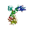
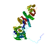
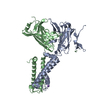
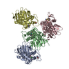
Crystal structure of NtrC-like two-domain protein (RER070207001320) from Eubacterium rectale at 2.10 A resolution
Components
NtrC-like two-domain protein
Keywords
structural genomics / unknown function / RER070207001320 / NtrC-like two-domain protein / Joint Center for Structural Genomics / JCSG / Protein Structure Initiative / PSI-2
Type: MARMOSAIC 325 mm CCD / Detector: CCD / Date: Nov 13, 2008 / Details: Flat mirror (vertical focusing)
Radiation
Monochromator: Single crystal Si(111) bent monochromator (horizontal focusing) Protocol: MAD / Monochromatic (M) / Laue (L): M / Scattering type: x-ray
Radiation wavelength
ID
Wavelength (Å)
Relative weight
1
0.91837
1
2
0.97908
1
3
0.97845
1
Reflection
Resolution: 2.1→29.161 Å / Num. obs: 30416 / % possible obs: 99.8 % / Redundancy: 3.2 % / Biso Wilson estimate: 36.83 Å2 / Rmerge(I) obs: 0.085 / Rsym value: 0.085 / Net I/σ(I): 5.832
Reflection shell
Diffraction-ID: 1
Resolution (Å)
Redundancy (%)
Rmerge(I) obs
Mean I/σ(I) obs
Num. measured all
Num. unique all
Rsym value
% possible all
2.1-2.15
3.2
0.627
1.2
7318
2258
0.627
100
2.15-2.21
3.2
0.534
1.4
7052
2179
0.534
100
2.21-2.28
3.2
0.429
1.7
6894
2126
0.429
100
2.28-2.35
3.2
0.373
2
6704
2067
0.373
100
2.35-2.42
3.3
0.318
2.3
6562
2016
0.318
100
2.42-2.51
3.2
0.254
2.9
6216
1919
0.254
100
2.51-2.6
3.2
0.211
3.4
6070
1868
0.211
100
2.6-2.71
3.2
0.185
3.9
5801
1791
0.185
100
2.71-2.83
3.2
0.154
4.5
5618
1732
0.154
100
2.83-2.97
3.2
0.116
6.1
5354
1655
0.116
100
2.97-3.13
3.2
0.1
6.6
5100
1579
0.1
100
3.13-3.32
3.2
0.081
7.8
4818
1488
0.081
100
3.32-3.55
3.2
0.072
8.6
4479
1384
0.072
99.9
3.55-3.83
3.2
0.062
9.8
4278
1319
0.062
99.8
3.83-4.2
3.2
0.057
10.5
3911
1208
0.057
99.7
4.2-4.7
3.2
0.051
11.5
3492
1076
0.051
99.6
4.7-5.42
3.2
0.054
11.5
3094
959
0.054
99.5
5.42-6.64
3.2
0.066
9.2
2585
819
0.066
99.3
6.64-9.39
3.2
0.059
9.7
1998
632
0.059
98.9
9.39-29.16
3.1
0.049
12.6
1042
341
0.049
95
-
Phasing
Phasing
Method: MAD
-
Processing
Software
Name
Version
Classification
NB
REFMAC
5.5.0053
refinement
PHENIX
refinement
SHELX
phasing
MolProbity
3beta29
modelbuilding
SCALA
3.2.5
datascaling
PDB_EXTRACT
3.006
dataextraction
MOSFLM
datareduction
SHELXD
phasing
autoSHARP
phasing
Refinement
Method to determine structure: MAD / Resolution: 2.1→29.161 Å / Cor.coef. Fo:Fc: 0.96 / Cor.coef. Fo:Fc free: 0.948 / Occupancy max: 1 / Occupancy min: 0.3 / SU B: 8.899 / SU ML: 0.112 / TLS residual ADP flag: LIKELY RESIDUAL / Cross valid method: THROUGHOUT / σ(F): 0 / ESU R: 0.171 / ESU R Free: 0.15 Stereochemistry target values: MAXIMUM LIKELIHOOD WITH PHASES Details: 1. HYDROGENS HAVE BEEN ADDED IN THE RIDING POSITIONS. 2. ATOM RECORDS CONTAIN RESIDUAL B FACTORS ONLY. 3. ATP AND MG ARE MODELED BASED ON DENSITY AND STRUCTURAL HOMOLOGS. PEG 400 (2PE), ...Details: 1. HYDROGENS HAVE BEEN ADDED IN THE RIDING POSITIONS. 2. ATOM RECORDS CONTAIN RESIDUAL B FACTORS ONLY. 3. ATP AND MG ARE MODELED BASED ON DENSITY AND STRUCTURAL HOMOLOGS. PEG 400 (2PE), SULFATE (SO4), CHLORIDE(CL), AND GLYCEROL (GOL) MODELED ARE PRESENT IN CRYO/CRYSTALLIZATION CONDITIONS
Rfactor
Num. reflection
% reflection
Selection details
Rfree
0.218
1535
5 %
RANDOM
Rwork
0.19
-
-
-
obs
0.191
30416
99.78 %
-
Solvent computation
Ion probe radii: 0.8 Å / Shrinkage radii: 0.8 Å / VDW probe radii: 1.2 Å / Solvent model: MASK
In the structure databanks used in Yorodumi, some data are registered as the other names, "COVID-19 virus" and "2019-nCoV". Here are the details of the virus and the list of structure data.
Jan 31, 2019. EMDB accession codes are about to change! (news from PDBe EMDB page)
EMDB accession codes are about to change! (news from PDBe EMDB page)
The allocation of 4 digits for EMDB accession codes will soon come to an end. Whilst these codes will remain in use, new EMDB accession codes will include an additional digit and will expand incrementally as the available range of codes is exhausted. The current 4-digit format prefixed with “EMD-” (i.e. EMD-XXXX) will advance to a 5-digit format (i.e. EMD-XXXXX), and so on. It is currently estimated that the 4-digit codes will be depleted around Spring 2019, at which point the 5-digit format will come into force.
The EM Navigator/Yorodumi systems omit the EMD- prefix.
Related info.:Q: What is EMD? / ID/Accession-code notation in Yorodumi/EM Navigator
Yorodumi is a browser for structure data from EMDB, PDB, SASBDB, etc.
This page is also the successor to EM Navigator detail page, and also detail information page/front-end page for Omokage search.
The word "yorodu" (or yorozu) is an old Japanese word meaning "ten thousand". "mi" (miru) is to see.
Related info.:EMDB / PDB / SASBDB / Comparison of 3 databanks / Yorodumi Search / Aug 31, 2016. New EM Navigator & Yorodumi / Yorodumi Papers / Jmol/JSmol / Function and homology information / Changes in new EM Navigator and Yorodumi
 Movie
Movie Controller

 Yorodumi
 Open data
 Basic information
 Components
 Keywords
 Function and homology information
 Eubacterium rectale (bacteria)
 X-RAY DIFFRACTION /
  Authors
 Citation
 Structure visualization
 Downloads & links
 Other downloads


 PDBj
PDBj Assembly
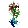


 Mass: 507.181 Da / Num. of mol.: 1 / Source method: obtained synthetically / Formula: C10H16N5O13P3 / Comment: ATP, energy-carrying molecule*YM
 Mass: 24.305 Da / Num. of mol.: 1 / Source method: obtained synthetically / Formula: Mg
 Mass: 35.453 Da / Num. of mol.: 1 / Source method: obtained synthetically / Formula: Cl
 Mass: 414.488 Da / Num. of mol.: 2 / Source method: obtained synthetically / Formula: C18H38O10 / Comment: precipitant*YM
 Mass: 92.094 Da / Num. of mol.: 2 / Source method: obtained synthetically / Formula: C3H8O3
 Mass: 96.063 Da / Num. of mol.: 1 / Source method: obtained synthetically / Formula: SO4
 Sample preparation
 / Beamline: BL11-1 / Wavelength: 0.91837,0.97908,0.97845
 Processing