 Movie
Movie Controller

[English] 日本語
 Yorodumi
- PDB-2ckq: Crystal structure of Human Choline Kinase alpha 2 in complex with... -
+ Open data

- Basic information
| Entry | Database: PDB / ID: 2ckq | ||||||
|---|---|---|---|---|---|---|---|
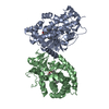
| Title | Crystal structure of Human Choline Kinase alpha 2 in complex with Phosphocholine | ||||||
 Components Components | CHOLINE KINASE ALPHA | ||||||
 Keywords Keywords | TRANSFERASE / PHOSPHATIDYLCHOLINE / PHOSPHOLIPID SYNTHESIS | ||||||
| Function / homology |  Function and homology information Function and homology informationethanolamine kinase / choline kinase / Synthesis of PE / ethanolamine kinase activity / choline kinase activity / CDP-choline pathway / lipid droplet disassembly / phosphatidylethanolamine biosynthetic process / phosphatidylcholine biosynthetic process / cholinesterase activity ...ethanolamine kinase / choline kinase / Synthesis of PE / ethanolamine kinase activity / choline kinase activity / CDP-choline pathway / lipid droplet disassembly / phosphatidylethanolamine biosynthetic process / phosphatidylcholine biosynthetic process / cholinesterase activity / lipid transport / Synthesis of PC / cellular response to glucose starvation / lipid droplet / lipid metabolic process / protein tyrosine kinase activity / protein homodimerization activity / ATP binding / cytoplasm / cytosol Similarity search - Function | ||||||
| Biological species |  HOMO SAPIENS (human) HOMO SAPIENS (human) | ||||||
| Method |  X-RAY DIFFRACTION / SYNCHROTRON / MOLECULAR REPLACEMENT / Resolution: 2.4 Å X-RAY DIFFRACTION / SYNCHROTRON / MOLECULAR REPLACEMENT / Resolution: 2.4 Å | ||||||
 Authors Authors | Malito, E. / Lavie, A. | ||||||
 Citation Citation | Journal: J.Mol.Biol. / Year: 2006 Title: Elucidation of Human Choline Kinase Crystal Structures in Complex with the Products Adp or Phosphocholine. Authors: Malito, E. / Sekulic, N. / Too, W.C. / Konrad, M. / Lavie, A. | ||||||
| History |
|
- Structure visualization
| Structure viewer | Molecule: MolmilJmol/JSmol |
|---|
- Downloads & links
-Download
| PDBx/mmCIF format | 2ckq.cif.gz | 153.7 KB | Display | PDBx/mmCIF format |
|---|---|---|---|---|
| PDB format | pdb2ckq.ent.gz | 118.9 KB | Display | PDB format |
| PDBx/mmJSON format | 2ckq.json.gz | Tree view | PDBx/mmJSON format | |
| Others |  Other downloads Other downloads |
-Validation report
| Arichive directory | https://data.pdbj.org/pub/pdb/validation_reports/ck/2ckqftp://data.pdbj.org/pub/pdb/validation_reports/ck/2ckq | HTTPS FTP |
|---|
-Related structure data
| Related structure data |  2ckoC  2ckpC  1nw1S C: citing same article ( S: Starting model for refinement |
|---|---|
| Similar structure data |

-Links
 PDBj
PDBj

- Assembly
| Deposited unit | 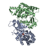
| ||||||||
|---|---|---|---|---|---|---|---|---|---|
| 1 |
| ||||||||
| Unit cell |
|
-Components
| #1: Protein | Mass: 45376.262 Da / Num. of mol.: 2 / Fragment: SPLICE ISOFORM 2, RESIDUES 50-439 Source method: isolated from a genetically manipulated source Source: (gene. exp.) HOMO SAPIENS (human) / Production host:  References: UniProt: P35790, choline kinase, ethanolamine kinase #2: Chemical |   Mass: 184.151 Da / Num. of mol.: 2 / Source method: obtained synthetically / Formula: C5H15NO4P Mass: 184.151 Da / Num. of mol.: 2 / Source method: obtained synthetically / Formula: C5H15NO4P#3: Water | ChemComp-HOH / |  Mass: 18.015 Da / Num. of mol.: 109 / Source method: isolated from a natural source / Formula: H2O Mass: 18.015 Da / Num. of mol.: 109 / Source method: isolated from a natural source / Formula: H2OSequence details | THE SEQUENCE DESCRIBES THE HUMAN CHOLINE KINASE ISOFORM ALFA-2, ACCORDING TO AYOAMA ET AL., 2000. ...THE SEQUENCE DESCRIBES THE HUMAN CHOLINE KINASE ISOFORM ALFA-2, ACCORDING TO AYOAMA ET AL., 2000. THE SEQUENCE FROM OUR CONSTRUCT LACKS THE FIRST N-TERMINAL 49 RESIDUES. THE SEQUENCE BELOW CORRESPOND | |
|---|
-Experimental details
-Experiment
| Experiment | Method: X-RAY DIFFRACTION / Number of used crystals: 1 |
|---|
- Sample preparation
| Crystal | Density Matthews: 3.64 Å3/Da / Density % sol: 65.9 % |
|---|---|
| Crystal grow | pH: 7.5 / Details: 0.01 M MGCL2, 15% PEG 3350, 0.2 M NAF., pH 7.50 |
-Data collection
| Diffraction | Mean temperature: 100 K |
|---|---|
| Diffraction source | Source: SYNCHROTRON / Site: APS  / Beamline: 22-ID / Wavelength: 1 / Beamline: 22-ID / Wavelength: 1 |
| Detector | Type: MARRESEARCH / Detector: CCD / Date: Oct 12, 2005 |
| Radiation | Protocol: SINGLE WAVELENGTH / Monochromatic (M) / Laue (L): M / Scattering type: x-ray |
| Radiation wavelength | Wavelength: 1 Å / Relative weight: 1 |
| Reflection | Resolution: 2.4→15 Å / Num. obs: 46369 / % possible obs: 99.8 % / Observed criterion σ(I): 0 / Redundancy: 8.1 % / Rmerge(I) obs: 0.11 / Net I/σ(I): 13.9 |
| Reflection shell | Resolution: 2.4→2.5 Å / Redundancy: 6.1 % / Rmerge(I) obs: 0.57 / Mean I/σ(I) obs: 3.2 / % possible all: 99.1 |
- Processing
| Software |
| ||||||||||||||||||||||||||||||||||||||||||||||||||||||||||||||||||||||||||||||||||||||||||||||||||||||||||||||||||||||||||||||||||||||||||||||||||||||||||||||||||||||||||||||||||||||
|---|---|---|---|---|---|---|---|---|---|---|---|---|---|---|---|---|---|---|---|---|---|---|---|---|---|---|---|---|---|---|---|---|---|---|---|---|---|---|---|---|---|---|---|---|---|---|---|---|---|---|---|---|---|---|---|---|---|---|---|---|---|---|---|---|---|---|---|---|---|---|---|---|---|---|---|---|---|---|---|---|---|---|---|---|---|---|---|---|---|---|---|---|---|---|---|---|---|---|---|---|---|---|---|---|---|---|---|---|---|---|---|---|---|---|---|---|---|---|---|---|---|---|---|---|---|---|---|---|---|---|---|---|---|---|---|---|---|---|---|---|---|---|---|---|---|---|---|---|---|---|---|---|---|---|---|---|---|---|---|---|---|---|---|---|---|---|---|---|---|---|---|---|---|---|---|---|---|---|---|---|---|---|---|
| Refinement | Method to determine structure: MOLECULAR REPLACEMENT Starting model: PDB ENTRY 1NW1 Resolution: 2.4→15 Å / Cor.coef. Fo:Fc: 0.938 / Cor.coef. Fo:Fc free: 0.909 / SU B: 17.554 / SU ML: 0.198 / TLS residual ADP flag: UNVERIFIED / Cross valid method: THROUGHOUT / ESU R: 0.294 / ESU R Free: 0.238 / Stereochemistry target values: MAXIMUM LIKELIHOOD
| ||||||||||||||||||||||||||||||||||||||||||||||||||||||||||||||||||||||||||||||||||||||||||||||||||||||||||||||||||||||||||||||||||||||||||||||||||||||||||||||||||||||||||||||||||||||
| Solvent computation | Ion probe radii: 0.8 Å / Shrinkage radii: 0.8 Å / VDW probe radii: 1.2 Å / Solvent model: BABINET MODEL WITH MASK | ||||||||||||||||||||||||||||||||||||||||||||||||||||||||||||||||||||||||||||||||||||||||||||||||||||||||||||||||||||||||||||||||||||||||||||||||||||||||||||||||||||||||||||||||||||||
| Displacement parameters | Biso mean: 35.66 Å2
| ||||||||||||||||||||||||||||||||||||||||||||||||||||||||||||||||||||||||||||||||||||||||||||||||||||||||||||||||||||||||||||||||||||||||||||||||||||||||||||||||||||||||||||||||||||||
| Refinement step | Cycle: LAST / Resolution: 2.4→15 Å
| ||||||||||||||||||||||||||||||||||||||||||||||||||||||||||||||||||||||||||||||||||||||||||||||||||||||||||||||||||||||||||||||||||||||||||||||||||||||||||||||||||||||||||||||||||||||
| Refine LS restraints |
|