 Movie
Movie Controller

[English] 日本語
 Yorodumi
- PDB-6f85: Crystal structure of cytochrome P450 CYP260A1 (S276N) bound with ... -
+ Open data

- Basic information
| Entry | Database: PDB / ID: 6f85 | |||||||||
|---|---|---|---|---|---|---|---|---|---|---|
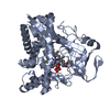
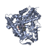
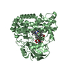
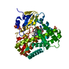
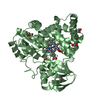
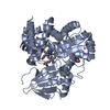
| Title | Crystal structure of cytochrome P450 CYP260A1 (S276N) bound with histidine | |||||||||
 Components Components | Cytochrome P450 CYP260A1 | |||||||||
 Keywords Keywords | OXIDOREDUCTASE / Bacterial Proteins / Sorangium cellulosum / Cytochrome P-450 Enzyme System / Cytochrome P450 / Hydroxylation / Heme / Oxidation-Reduction / Progesterone / Histidine / Biocatalysis | |||||||||
| Function / homology |  Function and homology information Function and homology informationC-19 steroid 1alpha-hydroxylase / oxidoreductase activity, acting on paired donors, with incorporation or reduction of molecular oxygen / monooxygenase activity / iron ion binding / heme binding Similarity search - Function | |||||||||
| Biological species |  Sorangium cellulosum (bacteria) Sorangium cellulosum (bacteria) | |||||||||
| Method |  X-RAY DIFFRACTION / SYNCHROTRON / MOLECULAR REPLACEMENT / Resolution: 2.05 Å X-RAY DIFFRACTION / SYNCHROTRON / MOLECULAR REPLACEMENT / Resolution: 2.05 Å | |||||||||
 Authors Authors | Jozwik, I.K. / Thunnissen, A.M.W.H. | |||||||||
| Funding support |  Germany, Germany,  Netherlands, 2items Netherlands, 2items
| |||||||||
 Citation Citation | Journal: ACS Chem. Biol. / Year: 2018 Title: Structure-Based Engineering of Steroidogenic CYP260A1 for Stereo- and Regioselective Hydroxylation of Progesterone. Authors: Khatri, Y. / Jozwik, I.K. / Ringle, M. / Ionescu, I.A. / Litzenburger, M. / Hutter, M.C. / Thunnissen, A.W.H. / Bernhardt, R. | |||||||||
| History |
|
- Structure visualization
| Structure viewer | Molecule: MolmilJmol/JSmol |
|---|
- Downloads & links
-Download
| PDBx/mmCIF format | 6f85.cif.gz | 175.2 KB | Display | PDBx/mmCIF format |
|---|---|---|---|---|
| PDB format | pdb6f85.ent.gz | 138.9 KB | Display | PDB format |
| PDBx/mmJSON format | 6f85.json.gz | Tree view | PDBx/mmJSON format | |
| Others |  Other downloads Other downloads |
-Validation report
| Arichive directory | https://data.pdbj.org/pub/pdb/validation_reports/f8/6f85ftp://data.pdbj.org/pub/pdb/validation_reports/f8/6f85 | HTTPS FTP |
|---|
-Related structure data
| Related structure data |  6f88C 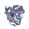 6f8aC 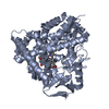 6f8cC 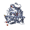 5livS S: Starting model for refinement C: citing same article ( |
|---|---|
| Similar structure data |


-Links
 PDBj
PDBj

- Assembly
| Deposited unit | 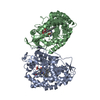
| ||||||||
|---|---|---|---|---|---|---|---|---|---|
| 1 | 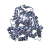
| ||||||||
| 2 | 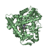
| ||||||||
| Unit cell |
|
-Components
| #1: Protein | Mass: 44524.922 Da / Num. of mol.: 2 / Mutation: S276N Source method: isolated from a genetically manipulated source Source: (gene. exp.) Sorangium cellulosum (strain So ce56) (bacteria)Strain: So ce56 / Gene: sce1588 / Plasmid: pET22b / Production host: References: UniProt: A9FDB7, Oxidoreductases; Acting on paired donors, with incorporation or reduction of molecular oxygen #2: Chemical |   Mass: 616.487 Da / Num. of mol.: 2 / Source method: obtained synthetically / Formula: C34H32FeN4O4 Mass: 616.487 Da / Num. of mol.: 2 / Source method: obtained synthetically / Formula: C34H32FeN4O4#3: Chemical |   Type: L-peptide linking / Mass: 156.162 Da / Num. of mol.: 2 / Source method: obtained synthetically / Formula: C6H10N3O2 Type: L-peptide linking / Mass: 156.162 Da / Num. of mol.: 2 / Source method: obtained synthetically / Formula: C6H10N3O2#4: Water | ChemComp-HOH / |  Mass: 18.015 Da / Num. of mol.: 294 / Source method: isolated from a natural source / Formula: H2O Mass: 18.015 Da / Num. of mol.: 294 / Source method: isolated from a natural source / Formula: H2O |
|---|
-Experimental details
-Experiment
| Experiment | Method: X-RAY DIFFRACTION / Number of used crystals: 1 |
|---|
- Sample preparation
| Crystal | Density Matthews: 2.15 Å3/Da / Density % sol: 42.68 % |
|---|---|
| Crystal grow | Temperature: 293 K / Method: vapor diffusion, hanging drop / pH: 6.5 Details: 0.1 M Bis-Tris propane pH 6.5, 14% (w/v) PEG 3350, 0.25 M NaNO3 |
-Data collection
| Diffraction | Mean temperature: 100 K | ||||||||||||||||||||||||
|---|---|---|---|---|---|---|---|---|---|---|---|---|---|---|---|---|---|---|---|---|---|---|---|---|---|
| Diffraction source | Source: SYNCHROTRON / Site: ESRF  / Beamline: ID29 / Wavelength: 0.97625 Å / Beamline: ID29 / Wavelength: 0.97625 Å | ||||||||||||||||||||||||
| Detector | Type: DECTRIS PILATUS 6M / Detector: PIXEL / Date: Nov 12, 2015 | ||||||||||||||||||||||||
| Radiation | Protocol: SINGLE WAVELENGTH / Monochromatic (M) / Laue (L): M / Scattering type: x-ray | ||||||||||||||||||||||||
| Radiation wavelength | Wavelength: 0.97625 Å / Relative weight: 1 | ||||||||||||||||||||||||
| Reflection | Resolution: 2.05→46.33 Å / Num. obs: 47060 / % possible obs: 99.2 % / Redundancy: 4.3 % / Biso Wilson estimate: 31.16 Å2 / CC1/2: 0.999 / Rmerge(I) obs: 0.047 / Rpim(I) all: 0.026 / Rrim(I) all: 0.054 / Net I/σ(I): 17.8 | ||||||||||||||||||||||||
| Reflection shell | Diffraction-ID: 1
|
- Processing
| Software |
| |||||||||||||||||||||||||||||||||||||||||||||||||||||||||||||||||||||||||||||||||||||||||||||||||||||||||||||||||||||||||||||||||||||||||||||||||||||||||||||||||||||||||||||||||||||||||||||||||||||||||||||||||||||||||
|---|---|---|---|---|---|---|---|---|---|---|---|---|---|---|---|---|---|---|---|---|---|---|---|---|---|---|---|---|---|---|---|---|---|---|---|---|---|---|---|---|---|---|---|---|---|---|---|---|---|---|---|---|---|---|---|---|---|---|---|---|---|---|---|---|---|---|---|---|---|---|---|---|---|---|---|---|---|---|---|---|---|---|---|---|---|---|---|---|---|---|---|---|---|---|---|---|---|---|---|---|---|---|---|---|---|---|---|---|---|---|---|---|---|---|---|---|---|---|---|---|---|---|---|---|---|---|---|---|---|---|---|---|---|---|---|---|---|---|---|---|---|---|---|---|---|---|---|---|---|---|---|---|---|---|---|---|---|---|---|---|---|---|---|---|---|---|---|---|---|---|---|---|---|---|---|---|---|---|---|---|---|---|---|---|---|---|---|---|---|---|---|---|---|---|---|---|---|---|---|---|---|---|---|---|---|---|---|---|---|---|---|---|---|---|---|---|---|---|
| Refinement | Method to determine structure: MOLECULAR REPLACEMENT Starting model: 5liv Resolution: 2.05→46.33 Å / SU ML: 0.2 / Cross valid method: FREE R-VALUE / σ(F): 0.03 / Phase error: 22.98
| |||||||||||||||||||||||||||||||||||||||||||||||||||||||||||||||||||||||||||||||||||||||||||||||||||||||||||||||||||||||||||||||||||||||||||||||||||||||||||||||||||||||||||||||||||||||||||||||||||||||||||||||||||||||||
| Solvent computation | Shrinkage radii: 0.9 Å / VDW probe radii: 1.11 Å | |||||||||||||||||||||||||||||||||||||||||||||||||||||||||||||||||||||||||||||||||||||||||||||||||||||||||||||||||||||||||||||||||||||||||||||||||||||||||||||||||||||||||||||||||||||||||||||||||||||||||||||||||||||||||
| Displacement parameters | Biso max: 99.08 Å2 / Biso mean: 36.4781 Å2 / Biso min: 18.18 Å2 | |||||||||||||||||||||||||||||||||||||||||||||||||||||||||||||||||||||||||||||||||||||||||||||||||||||||||||||||||||||||||||||||||||||||||||||||||||||||||||||||||||||||||||||||||||||||||||||||||||||||||||||||||||||||||
| Refinement step | Cycle: final / Resolution: 2.05→46.33 Å
| |||||||||||||||||||||||||||||||||||||||||||||||||||||||||||||||||||||||||||||||||||||||||||||||||||||||||||||||||||||||||||||||||||||||||||||||||||||||||||||||||||||||||||||||||||||||||||||||||||||||||||||||||||||||||
| Refine LS restraints |
| |||||||||||||||||||||||||||||||||||||||||||||||||||||||||||||||||||||||||||||||||||||||||||||||||||||||||||||||||||||||||||||||||||||||||||||||||||||||||||||||||||||||||||||||||||||||||||||||||||||||||||||||||||||||||
| LS refinement shell | Refine-ID: X-RAY DIFFRACTION / Rfactor Rfree error: 0 / Total num. of bins used: 30
|
