[English] 日本語
Yorodumi
- PDB-2c9x: Sulfite dehydrogenase from Starkeya Novella Y236F mutant -

+
Open data


ID or keywords:

Loading...

-
Basic information

Entry
Database: PDB / ID: 2c9x
TitleSulfite dehydrogenase from Starkeya Novella Y236F mutant
Components(SULFITE\:CYTOCHROME C OXIDOREDUCTASE SUBUNIT ...) x 2
KeywordsOXIDOREDUCTASE / SULFITE OXIDASE / MOLYBDOPTERIN / C-TYPE CYTOCHROME / HEME / MUTANT
Function / homology
Function and homology information


molybdenum ion binding / electron transfer activity / oxidoreductase activity / heme binding / metal ion binding
Similarity search - Function
Immunoglobulin-like - #650 / Sulfite Oxidase; Chain A, domain 2 / Oxidoreductase, molybdopterin-binding domain / Moybdenum cofactor oxidoreductase, dimerisation / Eukaryotic molybdopterin oxidoreductase / Mo-co oxidoreductase dimerisation domain / Oxidoreductase, molybdopterin-binding domain / Oxidoreductase molybdopterin binding domain / Oxidoreductase, molybdopterin-binding domain superfamily / Cytochrome c-like domain ...Immunoglobulin-like - #650 / Sulfite Oxidase; Chain A, domain 2 / Oxidoreductase, molybdopterin-binding domain / Moybdenum cofactor oxidoreductase, dimerisation / Eukaryotic molybdopterin oxidoreductase / Mo-co oxidoreductase dimerisation domain / Oxidoreductase, molybdopterin-binding domain / Oxidoreductase molybdopterin binding domain / Oxidoreductase, molybdopterin-binding domain superfamily / Cytochrome c-like domain / Cytochrome Bc1 Complex; Chain D, domain 2 / Cytochrome c-like domain superfamily / Twin arginine translocation (Tat) signal profile. / Twin-arginine translocation pathway, signal sequence / Immunoglobulin E-set / Alpha-Beta Complex / Immunoglobulin-like / Sandwich / Orthogonal Bundle / Mainly Beta / Mainly Alpha / Alpha Beta
Similarity search - Domain/homology
HEME C / (MOLYBDOPTERIN-S,S)-OXO-MOLYBDENUM / Sulfite:cytochrome c oxidoreductase subunit B / Sulfite:cytochrome c oxidoreductase subunit A
Similarity search - Component
Biological speciesTHIOBACILLUS NOVELLUS (bacteria)
MethodX-RAY DIFFRACTION / SYNCHROTRON / MOLECULAR REPLACEMENT / Resolution: 1.8 Å
AuthorsBailey, S. / Kappler, U. / Feng, C. / Honeychurch, M.J. / Bernhardt, P. / Tollin, G. / Enemark, J.
Citation
Journal: Biochemistry / Year: 2006
Title: Kinetic and Structural Evidence for the Importance of Tyr236 for the Integrity of the Mo Active Site in a Bacterial Sulfite Dehydrogenase.
Authors: Kappler, U. / Bailey, S. / Feng, C. / Honeychurch, M.J. / Hanson, G.R. / Bernhardt, P. / Tollin, G. / Enemark, J.
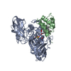
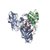
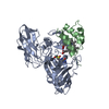
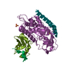
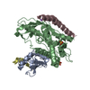
#1: Journal: J.Biol.Chem. / Year: 2005
Title: Molecular Basis of Intramolecular Electron Transfer in Sulfite-Oxidising Enzymes is Revealed by High Resolution Structure of a Heterodimeric Complex of the Catalytic Molybdopterin Subunit and ...Title: Molecular Basis of Intramolecular Electron Transfer in Sulfite-Oxidising Enzymes is Revealed by High Resolution Structure of a Heterodimeric Complex of the Catalytic Molybdopterin Subunit and a C-Type Cytochrome Subunit
Authors: Kappler, U. / Bailey, S.
History
DepositionDec 15, 2005Deposition site: PDBE / Processing site: PDBE
Revision 1.0Aug 16, 2006Provider: repository / Type: Initial release
Revision 1.1May 8, 2011Group: Version format compliance
Revision 1.2Jul 13, 2011Group: Version format compliance
Revision 1.3Dec 13, 2023Group: Data collection / Database references ...Data collection / Database references / Derived calculations / Other / Refinement description
Category: chem_comp_atom / chem_comp_bond ...chem_comp_atom / chem_comp_bond / database_2 / pdbx_database_status / pdbx_initial_refinement_model / struct_conn / struct_conn_type
Item: _database_2.pdbx_DOI / _database_2.pdbx_database_accession ..._database_2.pdbx_DOI / _database_2.pdbx_database_accession / _pdbx_database_status.status_code_sf / _struct_conn.conn_type_id / _struct_conn.id / _struct_conn.pdbx_dist_value / _struct_conn.pdbx_leaving_atom_flag / _struct_conn.ptnr1_auth_asym_id / _struct_conn.ptnr1_auth_comp_id / _struct_conn.ptnr1_auth_seq_id / _struct_conn.ptnr1_label_asym_id / _struct_conn.ptnr1_label_atom_id / _struct_conn.ptnr1_label_comp_id / _struct_conn.ptnr1_label_seq_id / _struct_conn.ptnr2_auth_asym_id / _struct_conn.ptnr2_auth_comp_id / _struct_conn.ptnr2_auth_seq_id / _struct_conn.ptnr2_label_asym_id / _struct_conn.ptnr2_label_atom_id / _struct_conn.ptnr2_label_comp_id / _struct_conn.ptnr2_label_seq_id / _struct_conn_type.id
Remark 700 SHEET THE SHEET STRUCTURE OF THIS MOLECULE IS BIFURCATED. IN ORDER TO REPRESENT THIS FEATURE IN ... SHEET THE SHEET STRUCTURE OF THIS MOLECULE IS BIFURCATED. IN ORDER TO REPRESENT THIS FEATURE IN THE SHEET RECORDS BELOW, TWO SHEETS ARE DEFINED.

-
Structure visualization

Structure viewerMolecule:
MolmilJmol/JSmol

Downloads & links

-
Assembly

Deposited unit
A: SULFITE\:CYTOCHROME C OXIDOREDUCTASE SUBUNIT A
B: SULFITE\:CYTOCHROME C OXIDOREDUCTASE SUBUNIT B
hetero molecules


Theoretical massNumber of molelcules
Total (without water)50,2435
Polymers49,0242
Non-polymers1,2203
Water7,584421
1


  • Idetical with deposited unit
  • defined by author&software
TypeNameSymmetry operationNumber
identity operation1_555x,y,z1
MethodPQS
Unit cell
Length a, b, c (Å)97.323, 93.214, 56.136
Angle α, β, γ (deg.)90.00, 90.00, 90.00
Int Tables number18
Space group name H-MP21212

-
Components

-
SULFITE\:CYTOCHROME C OXIDOREDUCTASE SUBUNIT ... , 2 types, 2 molecules AB

#1: Protein SULFITE\:CYTOCHROME C OXIDOREDUCTASE SUBUNIT A


Mass: 40178.719 Da / Num. of mol.: 1 / Mutation: YES
Source method: isolated from a genetically manipulated source
Source: (gene. exp.) THIOBACILLUS NOVELLUS (bacteria) / Production host: RHODOBACTER CAPSULATUS (bacteria) / References: UniProt: Q9LA16
#2: Protein SULFITE\:CYTOCHROME C OXIDOREDUCTASE SUBUNIT B


Mass: 8844.831 Da / Num. of mol.: 1
Source method: isolated from a genetically manipulated source
Source: (gene. exp.) THIOBACILLUS NOVELLUS (bacteria) / Production host: RHODOBACTER CAPSULATUS (bacteria) / References: UniProt: Q9LA15

-
Non-polymers , 4 types, 424 molecules

#3: Chemical ChemComp-MSS / (MOLYBDOPTERIN-S,S)-OXO-MOLYBDENUM


Mass: 505.275 Da / Num. of mol.: 1 / Source method: obtained synthetically / Formula: C10H12MoN5O7PS2
#4: Chemical ChemComp-SO4 / SULFATE ION / Sulfate


Mass: 96.063 Da / Num. of mol.: 1 / Source method: obtained synthetically / Formula: SO4
#5: Chemical ChemComp-HEC / HEME C / Heme C


Mass: 618.503 Da / Num. of mol.: 1 / Source method: obtained synthetically / Formula: C34H34FeN4O4
#6: Water ChemComp-HOH / water / Water


Mass: 18.015 Da / Num. of mol.: 421 / Source method: isolated from a natural source / Formula: H2O

-
Details

Compound detailsENGINEERED RESIDUE IN CHAIN A, TYR 268 TO PHE
Sequence detailsTHE TRANSLATED SEQUENCES FOR CHAINS A AND B INCLUDE SIGNAL PEPTIDES WHICH ARE NOT PART OF THE ...THE TRANSLATED SEQUENCES FOR CHAINS A AND B INCLUDE SIGNAL PEPTIDES WHICH ARE NOT PART OF THE ACTIVE, PROCESSED PROTEIN. THE SEQUENCES OF THE SIGNAL PEPTIDES ARE CHAIN A MLNNRRQILKSAGAAALGLGAGTLGWPGRHAFA CHAIN B MKRHNARTLAFSVVLGLSAAFAGSGLA

-
Experimental details

-
Experiment

ExperimentMethod: X-RAY DIFFRACTION / Number of used crystals: 1

-
Sample preparation

CrystalDensity Matthews: 2.6 Å3/Da / Density % sol: 46 %
Crystal growpH: 7.4
Details: 0.1M HEPES PH 7.4, 2.2M AMMONIUM SULFATE, 2% PEG200

-
Data collection

DiffractionMean temperature: 100 K
Diffraction sourceSource: SYNCHROTRON / Site: SRS / Beamline: PX14.2 / Wavelength: 0.98
DetectorType: ADSC CCD / Detector: CCD / Date: Nov 5, 2005
RadiationProtocol: SINGLE WAVELENGTH / Monochromatic (M) / Laue (L): M / Scattering type: x-ray
Radiation wavelengthWavelength: 0.98 Å / Relative weight: 1
ReflectionResolution: 1.8→50 Å / Num. obs: 46488 / % possible obs: 96.8 % / Observed criterion σ(I): 0 / Redundancy: 6.2 % / Rmerge(I) obs: 0.11 / Net I/σ(I): 14.5
Reflection shellResolution: 1.8→1.85 Å / Redundancy: 4 % / Rmerge(I) obs: 0.6 / Mean I/σ(I) obs: 2.2 / % possible all: 82.1

-
Processing

Software
NameVersionClassification
REFMAC5.2.0005refinement
MOSFLMdata reduction
SCALAdata scaling
RefinementMethod to determine structure: MOLECULAR REPLACEMENT
Starting model: PDB ENTRY 2BLF
Resolution: 1.8→20 Å / Cor.coef. Fo:Fc: 0.958 / Cor.coef. Fo:Fc free: 0.933 / SU B: 2.387 / SU ML: 0.075 / Cross valid method: THROUGHOUT / ESU R: 0.115 / ESU R Free: 0.116 / Stereochemistry target values: MAXIMUM LIKELIHOOD / Details: HYDROGENS HAVE BEEN ADDED IN THE RIDING POSITIONS.
RfactorNum. reflection% reflectionSelection details
Rfree0.209 2342 5 %RANDOM
Rwork0.166 ---
obs0.168 44052 96.6 %-
Solvent computationIon probe radii: 0.8 Å / Shrinkage radii: 0.8 Å / VDW probe radii: 1.2 Å / Solvent model: MASK
Displacement parametersBiso mean: 17.31 Å2
Baniso -1Baniso -2Baniso -3
1--0.32 Å20 Å20 Å2
2---0.48 Å20 Å2
3---0.8 Å2
Refinement stepCycle: LAST / Resolution: 1.8→20 Å
ProteinNucleic acidLigandSolventTotal
Num. atoms3454 0 74 421 3949
Refine LS restraints
Refine-IDTypeDev idealDev ideal targetNumber
X-RAY DIFFRACTIONr_bond_refined_d0.0130.0223629
X-RAY DIFFRACTIONr_bond_other_d
X-RAY DIFFRACTIONr_angle_refined_deg1.3912.014972
X-RAY DIFFRACTIONr_angle_other_deg
X-RAY DIFFRACTIONr_dihedral_angle_1_deg6.1965452
X-RAY DIFFRACTIONr_dihedral_angle_2_deg34.51124.305151
X-RAY DIFFRACTIONr_dihedral_angle_3_deg12.67715536
X-RAY DIFFRACTIONr_dihedral_angle_4_deg13.1791519
X-RAY DIFFRACTIONr_chiral_restr0.0890.2531
X-RAY DIFFRACTIONr_gen_planes_refined0.0060.022826
X-RAY DIFFRACTIONr_gen_planes_other
X-RAY DIFFRACTIONr_nbd_refined0.2110.21602
X-RAY DIFFRACTIONr_nbd_other
X-RAY DIFFRACTIONr_nbtor_refined0.3010.22476
X-RAY DIFFRACTIONr_nbtor_other
X-RAY DIFFRACTIONr_xyhbond_nbd_refined0.160.2322
X-RAY DIFFRACTIONr_xyhbond_nbd_other
X-RAY DIFFRACTIONr_metal_ion_refined
X-RAY DIFFRACTIONr_metal_ion_other
X-RAY DIFFRACTIONr_symmetry_vdw_refined0.1950.237
X-RAY DIFFRACTIONr_symmetry_vdw_other
X-RAY DIFFRACTIONr_symmetry_hbond_refined0.3410.228
X-RAY DIFFRACTIONr_symmetry_hbond_other
X-RAY DIFFRACTIONr_symmetry_metal_ion_refined
X-RAY DIFFRACTIONr_symmetry_metal_ion_other
X-RAY DIFFRACTIONr_mcbond_it3.24622322
X-RAY DIFFRACTIONr_mcbond_other
X-RAY DIFFRACTIONr_mcangle_it4.36543644
X-RAY DIFFRACTIONr_mcangle_other
X-RAY DIFFRACTIONr_scbond_it6.67751521
X-RAY DIFFRACTIONr_scbond_other
X-RAY DIFFRACTIONr_scangle_it9.125101326
X-RAY DIFFRACTIONr_scangle_other
X-RAY DIFFRACTIONr_long_range_B_refined
X-RAY DIFFRACTIONr_long_range_B_other
X-RAY DIFFRACTIONr_rigid_bond_restr
X-RAY DIFFRACTIONr_sphericity_free
X-RAY DIFFRACTIONr_sphericity_bonded
LS refinement shellResolution: 1.8→1.85 Å / Total num. of bins used: 20 /
RfactorNum. reflection
Rfree0.349 130
Rwork0.243 2538

+
About Yorodumi

-
News

-
Feb 9, 2022. New format data for meta-information of EMDB entries

New format data for meta-information of EMDB entries

  • Version 3 of the EMDB header file is now the official format.
  • The previous official version 1.9 will be removed from the archive.

Related info.:EMDB header

External links:wwPDB to switch to version 3 of the EMDB data model

-
Aug 12, 2020. Covid-19 info

Covid-19 info

URL: https://pdbj.org/emnavi/covid19.php

New page: Covid-19 featured information page in EM Navigator.

Related info.:Covid-19 info / Mar 5, 2020. Novel coronavirus structure data

+
Mar 5, 2020. Novel coronavirus structure data

Novel coronavirus structure data

Related info.:Yorodumi Speices / Aug 12, 2020. Covid-19 info

External links:COVID-19 featured content - PDBj / Molecule of the Month (242):Coronavirus Proteases

+
Jan 31, 2019. EMDB accession codes are about to change! (news from PDBe EMDB page)

EMDB accession codes are about to change! (news from PDBe EMDB page)

  • The allocation of 4 digits for EMDB accession codes will soon come to an end. Whilst these codes will remain in use, new EMDB accession codes will include an additional digit and will expand incrementally as the available range of codes is exhausted. The current 4-digit format prefixed with “EMD-” (i.e. EMD-XXXX) will advance to a 5-digit format (i.e. EMD-XXXXX), and so on. It is currently estimated that the 4-digit codes will be depleted around Spring 2019, at which point the 5-digit format will come into force.
  • The EM Navigator/Yorodumi systems omit the EMD- prefix.

Related info.:Q: What is EMD? / ID/Accession-code notation in Yorodumi/EM Navigator

External links:EMDB Accession Codes are Changing Soon! / Contact to PDBj

+
Jul 12, 2017. Major update of PDB

Major update of PDB

  • wwPDB released updated PDB data conforming to the new PDBx/mmCIF dictionary.
  • This is a major update changing the version number from 4 to 5, and with Remediation, in which all the entries are updated.
  • In this update, many items about electron microscopy experimental information are reorganized (e.g. em_software).
  • Now, EM Navigator and Yorodumi are based on the updated data.

External links:wwPDB Remediation / Enriched Model Files Conforming to OneDep Data Standards Now Available in the PDB FTP Archive

-
Yorodumi

Thousand views of thousand structures

  • Yorodumi is a browser for structure data from EMDB, PDB, SASBDB, etc.
  • This page is also the successor to EM Navigator detail page, and also detail information page/front-end page for Omokage search.
  • The word "yorodu" (or yorozu) is an old Japanese word meaning "ten thousand". "mi" (miru) is to see.

Related info.:EMDB / PDB / SASBDB / Comparison of 3 databanks / Yorodumi Search / Aug 31, 2016. New EM Navigator & Yorodumi / Yorodumi Papers / Jmol/JSmol / Function and homology information / Changes in new EM Navigator and Yorodumi

Read more