 Movie
Movie Controller


+ Open data

- Basic information
| Entry | Database: PDB / ID: 2ca4 | ||||||
|---|---|---|---|---|---|---|---|
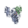
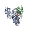
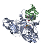
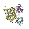
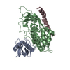
| Title | Sulfite dehydrogenase from Starkeya Novella mutant | ||||||
 Components Components |
| ||||||
 Keywords Keywords | OXIDOREDUCTASE / SULFITE OXIDASE / MOLYBDOPTERIN / C-TYPE CYTOCHROME / HEME / MUTANT | ||||||
| Function / homology |  Function and homology information Function and homology informationsulfite oxidase activity / sulfur compound metabolic process / molybdenum ion binding / molybdopterin cofactor binding / electron transfer activity / heme binding / metal ion binding Similarity search - Function | ||||||
| Biological species |  STARKEYA NOVELLA (bacteria) STARKEYA NOVELLA (bacteria) | ||||||
| Method |  X-RAY DIFFRACTION / SYNCHROTRON / MOLECULAR REPLACEMENT / Resolution: 2.1 Å X-RAY DIFFRACTION / SYNCHROTRON / MOLECULAR REPLACEMENT / Resolution: 2.1 Å | ||||||
 Authors Authors | Bailey, S. / Kappler, U. | ||||||
 Citation Citation | Journal: J.Biol.Chem. / Year: 2009 Title: Molecular Basis for Enzymatic Sulfite Oxidation: How Three Conserved Active Site Residues Shape Enzyme Activity. Authors: Bailey, S. / Rapson, T. / Johnson-Winters, K. / Astashkin, A.V. / Enemark, J.H. / Kappler, U. #1: Journal: J.Biol.Chem. / Year: 2005Title: Molecular Basis of Intramolecular Electron Transfer in Sulfite-Oxidizing Enzymes is Revealed by High Resolution Structure of a Heterodimeric Complex of the Catalytic Molybdopterin Subunit and ...Title: Molecular Basis of Intramolecular Electron Transfer in Sulfite-Oxidizing Enzymes is Revealed by High Resolution Structure of a Heterodimeric Complex of the Catalytic Molybdopterin Subunit and a C-Type Cytochrome Subunit Authors: Kappler, U. / Bailey, S. | ||||||
| History |
| ||||||
| Remark 700 | SHEET THE SHEET STRUCTURE OF THIS MOLECULE IS BIFURCATED. IN ORDER TO REPRESENT THIS FEATURE IN ... SHEET THE SHEET STRUCTURE OF THIS MOLECULE IS BIFURCATED. IN ORDER TO REPRESENT THIS FEATURE IN THE SHEET RECORDS BELOW, TWO SHEETS ARE DEFINED. |
- Structure visualization
| Structure viewer | Molecule: MolmilJmol/JSmol |
|---|
- Downloads & links
-Download
| PDBx/mmCIF format | 2ca4.cif.gz | 112.1 KB | Display | PDBx/mmCIF format |
|---|---|---|---|---|
| PDB format | pdb2ca4.ent.gz | 84.7 KB | Display | PDB format |
| PDBx/mmJSON format | 2ca4.json.gz | Tree view | PDBx/mmJSON format | |
| Others |  Other downloads Other downloads |
-Validation report
| Arichive directory | https://data.pdbj.org/pub/pdb/validation_reports/ca/2ca4ftp://data.pdbj.org/pub/pdb/validation_reports/ca/2ca4 | HTTPS FTP |
|---|
-Related structure data
| Related structure data | 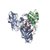 2ca3C  2blfS S: Starting model for refinement C: citing same article ( |
|---|---|
| Similar structure data |


-Links
 PDBj
PDBj


- Assembly
| Deposited unit | 
| ||||||||
|---|---|---|---|---|---|---|---|---|---|
| 1 |
| ||||||||
| Unit cell |
|
-Components
| #1: Protein | Mass: 40127.648 Da / Num. of mol.: 1 / Mutation: YES Source method: isolated from a genetically manipulated source Source: (gene. exp.) STARKEYA NOVELLA (bacteria) / Production host: RHODOBACTER CAPSULATUS (bacteria) / References: UniProt: Q9LA16 | ||||
|---|---|---|---|---|---|
| #2: Protein | Mass: 8844.831 Da / Num. of mol.: 1 Source method: isolated from a genetically manipulated source Source: (gene. exp.) STARKEYA NOVELLA (bacteria) / Production host: RHODOBACTER CAPSULATUS (bacteria) / References: UniProt: Q9LA15 | ||||
| #3: Chemical | ChemComp-MSS / (  Mass: 505.275 Da / Num. of mol.: 1 / Source method: obtained synthetically / Formula: C10H12MoN5O7PS2 Mass: 505.275 Da / Num. of mol.: 1 / Source method: obtained synthetically / Formula: C10H12MoN5O7PS2 | ||||
| #4: Chemical | ChemComp-HEC /   Mass: 618.503 Da / Num. of mol.: 1 / Source method: obtained synthetically / Formula: C34H34FeN4O4 Mass: 618.503 Da / Num. of mol.: 1 / Source method: obtained synthetically / Formula: C34H34FeN4O4 | ||||
| #5: Water | ChemComp-HOH /  Mass: 18.015 Da / Num. of mol.: 379 / Source method: isolated from a natural source / Formula: H2O Mass: 18.015 Da / Num. of mol.: 379 / Source method: isolated from a natural source / Formula: H2O | ||||
| Compound details | ENGINEERED| Has protein modification | Y | Sequence details | THE TRANSLATED SEQUENCES FOR CHAINS A AND B INCLUDE SIGNAL PEPTIDES WHICH ARE NOT PART OF THE ...THE TRANSLATED | |
-Experimental details
-Experiment
| Experiment | Method: X-RAY DIFFRACTION / Number of used crystals: 1 |
|---|
- Sample preparation
| Crystal | Density Matthews: 2.6 Å3/Da / Density % sol: 46 % |
|---|---|
| Crystal grow | pH: 7.4 Details: 0.1M HEPES PH 7.4, 2.2 M AMMONIUM SULFATE, 2% PEG 200 |
-Data collection
| Diffraction | Mean temperature: 100 K |
|---|---|
| Diffraction source | Source: SYNCHROTRON / Site: SRS  / Beamline: PX10.1 / Wavelength: 1.074 / Beamline: PX10.1 / Wavelength: 1.074 |
| Detector | Type: MARRESEARCH / Detector: CCD / Date: Nov 23, 2005 |
| Radiation | Protocol: SINGLE WAVELENGTH / Monochromatic (M) / Laue (L): M / Scattering type: x-ray |
| Radiation wavelength | Wavelength: 1.074 Å / Relative weight: 1 |
| Reflection | Resolution: 2.1→30 Å / Num. obs: 27649 / % possible obs: 93 % / Observed criterion σ(I): 0 / Redundancy: 3.4 % / Rmerge(I) obs: 0.1 / Net I/σ(I): 10.6 |
| Reflection shell | Resolution: 2.1→2.21 Å / Redundancy: 2 % / Rmerge(I) obs: 0.42 / Mean I/σ(I) obs: 2.1 / % possible all: 83.8 |
- Processing
| Software |
| ||||||||||||||||||||||||||||||||||||||||||||||||||||||||||||||||||||||||||||||||||||||||||||||||||||||||||||||||||||||||||||||||||||||||||||||||||||||||||||||||||||||||||||||||||||||
|---|---|---|---|---|---|---|---|---|---|---|---|---|---|---|---|---|---|---|---|---|---|---|---|---|---|---|---|---|---|---|---|---|---|---|---|---|---|---|---|---|---|---|---|---|---|---|---|---|---|---|---|---|---|---|---|---|---|---|---|---|---|---|---|---|---|---|---|---|---|---|---|---|---|---|---|---|---|---|---|---|---|---|---|---|---|---|---|---|---|---|---|---|---|---|---|---|---|---|---|---|---|---|---|---|---|---|---|---|---|---|---|---|---|---|---|---|---|---|---|---|---|---|---|---|---|---|---|---|---|---|---|---|---|---|---|---|---|---|---|---|---|---|---|---|---|---|---|---|---|---|---|---|---|---|---|---|---|---|---|---|---|---|---|---|---|---|---|---|---|---|---|---|---|---|---|---|---|---|---|---|---|---|---|
| Refinement | Method to determine structure: MOLECULAR REPLACEMENT Starting model: PDB ENTRY 2BLF Resolution: 2.1→20 Å / Cor.coef. Fo:Fc: 0.968 / Cor.coef. Fo:Fc free: 0.94 / SU B: 4.337 / SU ML: 0.113 / Cross valid method: THROUGHOUT / ESU R: 0.197 / ESU R Free: 0.169 / Stereochemistry target values: MAXIMUM LIKELIHOOD / Details: HYDROGENS HAVE BEEN ADDED IN THE RIDING POSITIONS.
| ||||||||||||||||||||||||||||||||||||||||||||||||||||||||||||||||||||||||||||||||||||||||||||||||||||||||||||||||||||||||||||||||||||||||||||||||||||||||||||||||||||||||||||||||||||||
| Solvent computation | Ion probe radii: 0.8 Å / Shrinkage radii: 0.8 Å / VDW probe radii: 1.2 Å / Solvent model: MASK | ||||||||||||||||||||||||||||||||||||||||||||||||||||||||||||||||||||||||||||||||||||||||||||||||||||||||||||||||||||||||||||||||||||||||||||||||||||||||||||||||||||||||||||||||||||||
| Displacement parameters | Biso mean: 23.1 Å2
| ||||||||||||||||||||||||||||||||||||||||||||||||||||||||||||||||||||||||||||||||||||||||||||||||||||||||||||||||||||||||||||||||||||||||||||||||||||||||||||||||||||||||||||||||||||||
| Refinement step | Cycle: LAST / Resolution: 2.1→20 Å
| ||||||||||||||||||||||||||||||||||||||||||||||||||||||||||||||||||||||||||||||||||||||||||||||||||||||||||||||||||||||||||||||||||||||||||||||||||||||||||||||||||||||||||||||||||||||
| Refine LS restraints |
|