 Movie
Movie Controller

[English] 日本語
 Yorodumi
- PDB-2c8c: Structure of the ARTT motif Q212A mutant C3bot1 Exoenzyme (NAD-bo... -
+ Open data

- Basic information
| Entry | Database: PDB / ID: 2c8c | ||||||
|---|---|---|---|---|---|---|---|
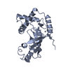
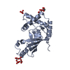
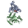
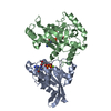
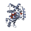
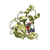
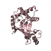
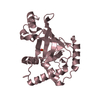
| Title | Structure of the ARTT motif Q212A mutant C3bot1 Exoenzyme (NAD-bound state, crystal form I) | ||||||
 Components Components | MONO-ADP-RIBOSYLTRANSFERASE C3 | ||||||
 Keywords Keywords | TRANSFERASE / C3 EXOENZYME / ARTT MOTIF / BACTERIAL TOXINS / GLYCOSYLTRANSFERASE | ||||||
| Function / homology |  Function and homology information Function and homology informationNAD+-protein mono-ADP-ribosyltransferase activity / Transferases; Glycosyltransferases; Pentosyltransferases / nucleotidyltransferase activity / extracellular region Similarity search - Function | ||||||
| Biological species |   CLOSTRIDIUM BOTULINUM (bacteria) CLOSTRIDIUM BOTULINUM (bacteria) | ||||||
| Method |  X-RAY DIFFRACTION / SYNCHROTRON / MOLECULAR REPLACEMENT / Resolution: 2.7 Å X-RAY DIFFRACTION / SYNCHROTRON / MOLECULAR REPLACEMENT / Resolution: 2.7 Å | ||||||
 Authors Authors | Stura, E.A. / Menetrey, J. / Flatau, G. / Boquet, P. / Menez, A. | ||||||
 Citation Citation | Journal: Protein Sci. / Year: 2008 Title: Structural Basis for the Nad-Hydrolysis Mechanism and the Artt-Loop Plasticity of C3 Exoenzymes. Authors: Menetrey, J. / Flatau, G. / Boquet, P. / Menez, A. / Stura, E.A. #1: Journal: J.Biol.Chem. / Year: 2002Title: Nad Binding Induces Conformational Changes in Rho Adp-Ribosylating Clostridium Botulinum C3 Exoenzyme. Authors: Menetrey, J. / Flatau, G. / Stura, E.A. / Charbonnier, J.B. / Gas, F. / Teulon, J.M. / Ledu, M.H. / Boquet, P. / Menez, A. | ||||||
| History |
|
- Structure visualization
| Structure viewer | Molecule: MolmilJmol/JSmol |
|---|
- Downloads & links
-Download
| PDBx/mmCIF format | 2c8c.cif.gz | 171.7 KB | Display | PDBx/mmCIF format |
|---|---|---|---|---|
| PDB format | pdb2c8c.ent.gz | 137 KB | Display | PDB format |
| PDBx/mmJSON format | 2c8c.json.gz | Tree view | PDBx/mmJSON format | |
| Others |  Other downloads Other downloads |
-Validation report
| Arichive directory | https://data.pdbj.org/pub/pdb/validation_reports/c8/2c8cftp://data.pdbj.org/pub/pdb/validation_reports/c8/2c8c | HTTPS FTP |
|---|
-Related structure data
| Related structure data |  2c89C  2c8aC  2c8bC  2c8dC  2c8eC  2c8fC  1gzfS S: Starting model for refinement C: citing same article ( |
|---|---|
| Similar structure data |


-Links
 PDBj
PDBj- Assembly
| Deposited unit | 
| ||||||||
|---|---|---|---|---|---|---|---|---|---|
| 1 | 
| ||||||||
| 2 | 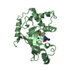
| ||||||||
| 3 | 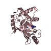
| ||||||||
| 4 | 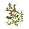
| ||||||||
| Unit cell |
|
-Components
| #1: Protein | Mass: 23534.918 Da / Num. of mol.: 4 / Mutation: YES Source method: isolated from a genetically manipulated source Source: (gene. exp.) CLOSTRIDIUM BOTULINUM (bacteria) / Production host: References: UniProt: P15879, UniProt: Q7M0L1*PLUS, Transferases; Glycosyltransferases; Pentosyltransferases #2: Chemical |   Mass: 663.425 Da / Num. of mol.: 3 / Source method: obtained synthetically / Formula: C21H27N7O14P2 / Comment: NAD*YM Mass: 663.425 Da / Num. of mol.: 3 / Source method: obtained synthetically / Formula: C21H27N7O14P2 / Comment: NAD*YM#3: Chemical | ChemComp-ADP / |   Mass: 427.201 Da / Num. of mol.: 1 / Source method: obtained synthetically / Formula: C10H15N5O10P2 / Comment: ADP, energy-carrying molecule*YM Mass: 427.201 Da / Num. of mol.: 1 / Source method: obtained synthetically / Formula: C10H15N5O10P2 / Comment: ADP, energy-carrying molecule*YMCompound details | ADP-RIBOSYLATES EUKARYOTIC RHO AND RAC PROTEINS ON AN ASPARAGINE RESIDUE ENGINEERED RESIDUE IN ...ADP-RIBOSYLATE | |
|---|
-Experimental details
-Experiment
| Experiment | Method: X-RAY DIFFRACTION |
|---|
- Sample preparation
| Crystal | Density Matthews: 2.57 Å3/Da / Density % sol: 51.83 % |
|---|---|
| Crystal grow | Details: 22.5% PEG 3350 W/W, 100 MM LI2SO4, 100 MM SODIUM CITRATE PH 3.0, 3-10% MPEG 550 V/V |
-Data collection
| Diffraction | Mean temperature: 100 K |
|---|---|
| Diffraction source | Source: SYNCHROTRON / Site: ESRF  / Beamline: ID14-3 / Wavelength: 0.933 / Beamline: ID14-3 / Wavelength: 0.933 |
| Detector | Type: ADSC CCD / Detector: CCD |
| Radiation | Protocol: SINGLE WAVELENGTH / Monochromatic (M) / Laue (L): M / Scattering type: x-ray |
| Radiation wavelength | Wavelength: 0.933 Å / Relative weight: 1 |
| Reflection | Resolution: 2.7→39.84 Å / Num. obs: 24821 / % possible obs: 95.8 % / Observed criterion σ(I): 2 / Redundancy: 4 % / Rmerge(I) obs: 0.09 / Net I/σ(I): 12.4 |
- Processing
| Software |
| ||||||||||||||||||||||||||||||||||||||||||||||||||||||||||||||||||||||||||||||||||||||||||||||||||||||||||||||||||||||||||||||||||||||||||||||||||||||||||||||||||||||||||||||||||||||
|---|---|---|---|---|---|---|---|---|---|---|---|---|---|---|---|---|---|---|---|---|---|---|---|---|---|---|---|---|---|---|---|---|---|---|---|---|---|---|---|---|---|---|---|---|---|---|---|---|---|---|---|---|---|---|---|---|---|---|---|---|---|---|---|---|---|---|---|---|---|---|---|---|---|---|---|---|---|---|---|---|---|---|---|---|---|---|---|---|---|---|---|---|---|---|---|---|---|---|---|---|---|---|---|---|---|---|---|---|---|---|---|---|---|---|---|---|---|---|---|---|---|---|---|---|---|---|---|---|---|---|---|---|---|---|---|---|---|---|---|---|---|---|---|---|---|---|---|---|---|---|---|---|---|---|---|---|---|---|---|---|---|---|---|---|---|---|---|---|---|---|---|---|---|---|---|---|---|---|---|---|---|---|---|
| Refinement | Method to determine structure: MOLECULAR REPLACEMENT Starting model: PDB ENTRY 1GZF Resolution: 2.7→39.84 Å / Cor.coef. Fo:Fc: 0.929 / Cor.coef. Fo:Fc free: 0.906 / SU B: 15.101 / SU ML: 0.301 / Cross valid method: THROUGHOUT / ESU R Free: 0.411 / Stereochemistry target values: MAXIMUM LIKELIHOOD / Details: HYDROGENS HAVE BEEN ADDED IN THE RIDING POSITIONS.
| ||||||||||||||||||||||||||||||||||||||||||||||||||||||||||||||||||||||||||||||||||||||||||||||||||||||||||||||||||||||||||||||||||||||||||||||||||||||||||||||||||||||||||||||||||||||
| Solvent computation | Ion probe radii: 0.8 Å / Shrinkage radii: 0.8 Å / VDW probe radii: 1.4 Å / Solvent model: BABINET MODEL WITH MASK | ||||||||||||||||||||||||||||||||||||||||||||||||||||||||||||||||||||||||||||||||||||||||||||||||||||||||||||||||||||||||||||||||||||||||||||||||||||||||||||||||||||||||||||||||||||||
| Displacement parameters | Biso mean: 56.76 Å2
| ||||||||||||||||||||||||||||||||||||||||||||||||||||||||||||||||||||||||||||||||||||||||||||||||||||||||||||||||||||||||||||||||||||||||||||||||||||||||||||||||||||||||||||||||||||||
| Refinement step | Cycle: LAST / Resolution: 2.7→39.84 Å
| ||||||||||||||||||||||||||||||||||||||||||||||||||||||||||||||||||||||||||||||||||||||||||||||||||||||||||||||||||||||||||||||||||||||||||||||||||||||||||||||||||||||||||||||||||||||
| Refine LS restraints |
|