| Entry | Database: PDB / ID: 2c7s
|
|---|
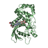
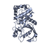
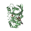
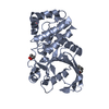
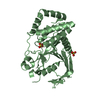
| Title | Crystal structure of human protein tyrosine phosphatase kappa at 1.95A resolution |
|---|
 Components Components | RECEPTOR-TYPE TYROSINE-PROTEIN PHOSPHATASE KAPPA |
|---|
 Keywords Keywords | HYDROLASE / RECEPTOR TYPE TYROSINE PHOSPHATASE KAPPA / PTPRK / GLYCOPROTEIN / IMMUNOGLOBULIN DOMAIN / PROTEIN PHOSPHATASE / RECEPTOR / TRANSMEMBRANE |
|---|
| Function / homology |  Function and homology information Function and homology information
transmembrane receptor protein tyrosine phosphatase activity / gamma-catenin binding / leading edge membrane / focal adhesion assembly / protein localization to cell surface / protein dephosphorylation / negative regulation of cell cycle / sperm head-tail coupling apparatus / negative regulation of keratinocyte proliferation / protein-tyrosine-phosphatase ...transmembrane receptor protein tyrosine phosphatase activity / gamma-catenin binding / leading edge membrane / focal adhesion assembly / protein localization to cell surface / protein dephosphorylation / negative regulation of cell cycle / sperm head-tail coupling apparatus / negative regulation of keratinocyte proliferation / protein-tyrosine-phosphatase / protein tyrosine phosphatase activity / transforming growth factor beta receptor signaling pathway / negative regulation of cell migration / adherens junction / cellular response to reactive oxygen species / centriole / EGFR downregulation / beta-catenin binding / neuron projection development / cell-cell junction / cellular response to UV / cell junction / cell migration / cell adhesion / negative regulation of cell population proliferation / negative regulation of DNA-templated transcription / protein kinase binding / cell surface / signal transduction / membrane / plasma membraneSimilarity search - Function : / Receptor-type tyrosine-protein phosphatase U Fn3 domain / MAM domain signature. / : / Domain in meprin, A5, receptor protein tyrosine phosphatase mu (and others) / MAM domain, meprin/A5/mu / MAM domain / MAM domain profile. / Protein tyrosine phosphatase superfamily / Protein-Tyrosine Phosphatase; Chain A ...: / Receptor-type tyrosine-protein phosphatase U Fn3 domain / MAM domain signature. / : / Domain in meprin, A5, receptor protein tyrosine phosphatase mu (and others) / MAM domain, meprin/A5/mu / MAM domain / MAM domain profile. / Protein tyrosine phosphatase superfamily / Protein-Tyrosine Phosphatase; Chain A / Protein tyrosine phosphatase, catalytic domain / PTP type protein phosphatase domain profile. / Protein-tyrosine phosphatase / Tyrosine-specific protein phosphatase, PTPase domain / Immunoglobulin I-set / Immunoglobulin I-set domain / Protein-tyrosine phosphatase, catalytic / Protein tyrosine phosphatase, catalytic domain motif / Tyrosine specific protein phosphatases active site. / Protein-tyrosine phosphatase, active site / Tyrosine specific protein phosphatases domain profile. / Tyrosine-specific protein phosphatases domain / Protein-tyrosine phosphatase-like / Fibronectin type III domain / Fibronectin type 3 domain / Fibronectin type-III domain profile. / Fibronectin type III / Fibronectin type III superfamily / Immunoglobulin subtype / Immunoglobulin / Concanavalin A-like lectin/glucanase domain superfamily / Ig-like domain profile. / Immunoglobulin-like domain / Immunoglobulin-like domain superfamily / Immunoglobulin-like fold / Alpha-Beta Complex / Alpha BetaSimilarity search - Domain/homology |
|---|
| Biological species |  HOMO SAPIENS (human) HOMO SAPIENS (human) |
|---|
| Method |  X-RAY DIFFRACTION / SYNCHROTRON / MOLECULAR REPLACEMENT / Resolution: 1.95 Å X-RAY DIFFRACTION / SYNCHROTRON / MOLECULAR REPLACEMENT / Resolution: 1.95 Å |
|---|
 Authors Authors | Debreczeni, J.E. / Ugochukwu, E. / Eswaran, J. / Barr, A. / Das, S. / Burgess, N. / Gileadi, O. / Longman, E. / von Delft, F. / Knapp, S. ...Debreczeni, J.E. / Ugochukwu, E. / Eswaran, J. / Barr, A. / Das, S. / Burgess, N. / Gileadi, O. / Longman, E. / von Delft, F. / Knapp, S. / Sundstron, M. / Arrowsmith, C. / Weigelt, J. / Edwards, A. |
|---|
 Citation Citation | Journal: Protein Sci. / Year: 2006
Title: The crystal structure of human receptor protein tyrosine phosphatase kappa phosphatase domain 1.
Authors: Eswaran, J. / Debreczeni, J.E. / Longman, E. / Barr, A.J. / Knapp, S. |
|---|
| History | | Deposition | Nov 28, 2005 | Deposition site: PDBE / Processing site: PDBE |
|---|
| Revision 1.0 | Jan 2, 2007 | Provider: repository / Type: Initial release |
|---|
| Revision 1.1 | Jul 13, 2011 | Group: Advisory / Version format compliance |
|---|
| Revision 1.2 | Jan 24, 2018 | Group: Structure summary / Category: audit_author / Item: _audit_author.name |
|---|
| Revision 1.3 | Feb 28, 2018 | Group: Database references / Source and taxonomy / Structure summary
Category: citation / citation_author ...citation / citation_author / entity_src_gen / struct
Item: _citation.page_last / _citation.pdbx_database_id_DOI ..._citation.page_last / _citation.pdbx_database_id_DOI / _citation.title / _citation_author.name / _entity_src_gen.pdbx_host_org_cell_line / _entity_src_gen.pdbx_host_org_ncbi_taxonomy_id / _entity_src_gen.pdbx_host_org_scientific_name / _entity_src_gen.pdbx_host_org_strain / _struct.title |
|---|
| Revision 1.4 | May 8, 2019 | Group: Data collection / Derived calculations / Experimental preparation
Category: exptl_crystal_grow / pdbx_struct_special_symmetry / Item: _exptl_crystal_grow.method |
|---|
| Revision 1.5 | Dec 13, 2023 | Group: Data collection / Database references ...Data collection / Database references / Other / Refinement description
Category: chem_comp_atom / chem_comp_bond ...chem_comp_atom / chem_comp_bond / database_2 / pdbx_database_status / pdbx_initial_refinement_model
Item: _database_2.pdbx_DOI / _database_2.pdbx_database_accession / _pdbx_database_status.status_code_sf |
|---|
| Revision 1.6 | Nov 13, 2024 | Group: Structure summary / Category: pdbx_entry_details / pdbx_modification_feature / Item: _pdbx_entry_details.has_protein_modification |
|---|
|
|---|
 Movie
Movie Controller

 Yorodumi
 Open data
 Basic information
 Structure visualization
 Downloads & links
 Other downloads


 PDBj
PDBj


 Assembly
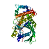


 Mass: 59.044 Da / Num. of mol.: 1 / Source method: obtained synthetically / Formula: C2H3O2
 Mass: 18.015 Da / Num. of mol.: 138 / Source method: isolated from a natural source / Formula: H2O
 Sample preparation
 / Beamline: X10SA / Wavelength: 0.978978
  Processing