 Movie
Movie Controller

[English] 日本語
 Yorodumi
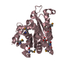
- PDB-2c3a: Structure of unliganded HSV gD reveals a mechanism for receptor- ... -
+ Open data

- Basic information
| Entry | Database: PDB / ID: 2c3a | |||||||||
|---|---|---|---|---|---|---|---|---|---|---|
| Title | Structure of unliganded HSV gD reveals a mechanism for receptor- mediated activation of virus entry | |||||||||
 Components Components | GLYCOPROTEIN D | |||||||||
 Keywords Keywords | VIRAL PROTEIN / VIRUS / HERPES SIMPLEX VIRUS / HSV-1 / GLYCOPROTEIN D | |||||||||
| Function / homology |  Function and homology information Function and homology informationhost cell Golgi apparatus / entry receptor-mediated virion attachment to host cell / receptor ligand activity / viral envelope / symbiont entry into host cell / virion membrane / membrane / metal ion binding Similarity search - Function | |||||||||
| Biological species |   HUMAN HERPESVIRUS 1 (Herpes simplex virus type 1) HUMAN HERPESVIRUS 1 (Herpes simplex virus type 1) | |||||||||
| Method |  X-RAY DIFFRACTION / SYNCHROTRON / MOLECULAR REPLACEMENT / Resolution: 2.5 Å X-RAY DIFFRACTION / SYNCHROTRON / MOLECULAR REPLACEMENT / Resolution: 2.5 Å | |||||||||
 Authors Authors | Krummenacher, C. / Supekar, V.M. / Whitbeck, J.C. / Lazear, E. / Connolly, S.A. / Eisenberg, R.J. / Cohen, G.H. / Wiley, D.C. / Carfi, A. | |||||||||
 Citation Citation | Journal: Embo J. / Year: 2005 Title: Structure of Unliganded Hsv Gd Reveals a Mechanism for Receptor-Mediated Activation of Virus Entry. Authors: Krummenacher, C. / Supekar, V.M. / Whitbeck, J.C. / Lazear, E. / Connolly, S.A. / Eisenberg, R.J. / Cohen, G.H. / Wiley, D.C. / Carfi, A. | |||||||||
| History |
| |||||||||
| Remark 700 | SHEET THE SHEET STRUCTURE OF THIS MOLECULE IS BIFURCATED. IN ORDER TO REPRESENT THIS FEATURE IN ... SHEET THE SHEET STRUCTURE OF THIS MOLECULE IS BIFURCATED. IN ORDER TO REPRESENT THIS FEATURE IN THE SHEET RECORDS BELOW, TWO SHEETS ARE DEFINED. |
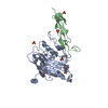
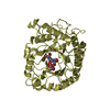
- Structure visualization
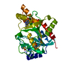
| Structure viewer | Molecule: MolmilJmol/JSmol |
|---|
- Downloads & links
-Download
| PDBx/mmCIF format | 2c3a.cif.gz | 120.1 KB | Display | PDBx/mmCIF format |
|---|---|---|---|---|
| PDB format | pdb2c3a.ent.gz | 91.6 KB | Display | PDB format |
| PDBx/mmJSON format | 2c3a.json.gz | Tree view | PDBx/mmJSON format | |
| Others |  Other downloads Other downloads |
-Validation report
| Arichive directory | https://data.pdbj.org/pub/pdb/validation_reports/c3/2c3aftp://data.pdbj.org/pub/pdb/validation_reports/c3/2c3a | HTTPS FTP |
|---|
-Related structure data
| Related structure data | 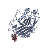 2c36C  1l2gS S: Starting model for refinement C: citing same article ( |
|---|---|
| Similar structure data |


-Links
 PDBj
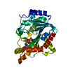
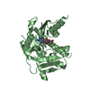
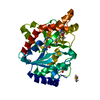
PDBj- Assembly
| Deposited unit | 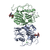
| ||||||||||||||||||
|---|---|---|---|---|---|---|---|---|---|---|---|---|---|---|---|---|---|---|---|
| 1 | 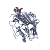
| ||||||||||||||||||
| 2 | 
| ||||||||||||||||||
| Unit cell |
| ||||||||||||||||||
| Noncrystallographic symmetry (NCS) | NCS domain:
NCS domain segments: Component-ID: 1 / Ens-ID: 1 / Beg auth comp-ID: PHE / Beg label comp-ID: PHE / End auth comp-ID: SER / End label comp-ID: SER / Refine code: 2 / Auth seq-ID: 100 - 200 / Label seq-ID: 79 - 179
NCS oper: (Code: given Matrix: (0.96045, 0.27667, 0.03155), Vector: |
-Components
-Protein / Sugars , 2 types, 4 molecules AB
| #1: Protein | Mass: 31743.059 Da / Num. of mol.: 2 / Mutation: YES Source method: isolated from a genetically manipulated source Details: N-ACETYL-D-GLUCOSAMINE LINKED TO ASN94 FOR BOTH CHAINS A AND B Source: (gene. exp.) HUMAN HERPESVIRUS 1 (Herpes simplex virus type 1)Strain: PATTON / Plasmid: PDL485 / Cell line (production host): SF9 / Production host:   SPODOPTERA FRUGIPERDA (fall armyworm) / References: UniProt: P57083 SPODOPTERA FRUGIPERDA (fall armyworm) / References: UniProt: P57083#2: Polysaccharide | Source method: isolated from a genetically manipulated source |
|---|
-Non-polymers , 4 types, 62 molecules 


| #3: Chemical |  Mass: 35.453 Da / Num. of mol.: 2 / Source method: obtained synthetically / Formula: Cl Mass: 35.453 Da / Num. of mol.: 2 / Source method: obtained synthetically / Formula: Cl#4: Chemical | ChemComp-ZN /  Mass: 65.409 Da / Num. of mol.: 5 / Source method: obtained synthetically / Formula: Zn Mass: 65.409 Da / Num. of mol.: 5 / Source method: obtained synthetically / Formula: Zn#5: Chemical |  Mass: 22.990 Da / Num. of mol.: 2 / Source method: obtained synthetically / Formula: Na Mass: 22.990 Da / Num. of mol.: 2 / Source method: obtained synthetically / Formula: Na#6: Water | ChemComp-HOH / | Mass: 18.015 Da / Num. of mol.: 53 / Source method: isolated from a natural source / Formula: H2O |
|---|
-Details
| Compound details | ENGINEERED RESIDUE IN CHAIN A, VAL 62 TO CYS ENGINEERED RESIDUE IN CHAIN A, ALA 327 TO CYS ...ENGINEERED |
|---|---|
| Has protein modification | Y |
-Experimental details
-Experiment
| Experiment | Method: X-RAY DIFFRACTION / Number of used crystals: 1 |
|---|
- Sample preparation
| Crystal | Density Matthews: 2.4 Å3/Da / Density % sol: 49 % |
|---|---|
| Crystal grow | pH: 4.6 / Details: 100MM NA ACETATE PH 4.6, 10MM ZNCL2, 24% PEG &K |
-Data collection
| Diffraction | Mean temperature: 100 K |
|---|---|
| Diffraction source | Source: SYNCHROTRON / Site: ESRF  / Beamline: ID14-3 / Wavelength: 0.931 / Beamline: ID14-3 / Wavelength: 0.931 |
| Detector | Type: MARRESEARCH / Detector: CCD / Date: Oct 20, 2004 |
| Radiation | Protocol: SINGLE WAVELENGTH / Monochromatic (M) / Laue (L): M / Scattering type: x-ray |
| Radiation wavelength | Wavelength: 0.931 Å / Relative weight: 1 |
| Reflection | Resolution: 2.5→50 Å / Num. obs: 23602 / % possible obs: 99.6 % / Observed criterion σ(I): -3 / Redundancy: 7.3 % / Rmerge(I) obs: 0.08 / Net I/σ(I): 9 |
| Reflection shell | Resolution: 2.5→2.59 Å / Redundancy: 7.1 % / Rmerge(I) obs: 0.34 / Mean I/σ(I) obs: 5.07 / % possible all: 98.2 |
- Processing
| Software |
| ||||||||||||||||||||||||||||||||||||||||||||||||||||||||||||||||||||||||||||||||||||||||||||||||||||||||||||||||||||||||||||||||||||||||||||||||||||||||||||||||||||||||||||||||||||||
|---|---|---|---|---|---|---|---|---|---|---|---|---|---|---|---|---|---|---|---|---|---|---|---|---|---|---|---|---|---|---|---|---|---|---|---|---|---|---|---|---|---|---|---|---|---|---|---|---|---|---|---|---|---|---|---|---|---|---|---|---|---|---|---|---|---|---|---|---|---|---|---|---|---|---|---|---|---|---|---|---|---|---|---|---|---|---|---|---|---|---|---|---|---|---|---|---|---|---|---|---|---|---|---|---|---|---|---|---|---|---|---|---|---|---|---|---|---|---|---|---|---|---|---|---|---|---|---|---|---|---|---|---|---|---|---|---|---|---|---|---|---|---|---|---|---|---|---|---|---|---|---|---|---|---|---|---|---|---|---|---|---|---|---|---|---|---|---|---|---|---|---|---|---|---|---|---|---|---|---|---|---|---|---|
| Refinement | Method to determine structure: MOLECULAR REPLACEMENT Starting model: PDB ENTRY 1L2G Resolution: 2.5→25 Å / Cor.coef. Fo:Fc: 0.928 / Cor.coef. Fo:Fc free: 0.896 / SU B: 8.386 / SU ML: 0.193 / Cross valid method: THROUGHOUT / ESU R: 0.517 / ESU R Free: 0.307 / Stereochemistry target values: MAXIMUM LIKELIHOOD Details: HYDROGENS HAVE BEEN ADDED IN THE RIDING POSITIONS. FIRST 22 RESIDUES DISORDERED FOR BOTH CHAINS A,B. RESIDUES 256-267 DISORDERED FOR BOTH CHAINS A,B
| ||||||||||||||||||||||||||||||||||||||||||||||||||||||||||||||||||||||||||||||||||||||||||||||||||||||||||||||||||||||||||||||||||||||||||||||||||||||||||||||||||||||||||||||||||||||
| Solvent computation | Ion probe radii: 0.8 Å / Shrinkage radii: 0.8 Å / VDW probe radii: 1.4 Å / Solvent model: BABINET MODEL WITH MASK | ||||||||||||||||||||||||||||||||||||||||||||||||||||||||||||||||||||||||||||||||||||||||||||||||||||||||||||||||||||||||||||||||||||||||||||||||||||||||||||||||||||||||||||||||||||||
| Displacement parameters | Biso mean: 39 Å2
| ||||||||||||||||||||||||||||||||||||||||||||||||||||||||||||||||||||||||||||||||||||||||||||||||||||||||||||||||||||||||||||||||||||||||||||||||||||||||||||||||||||||||||||||||||||||
| Refinement step | Cycle: LAST / Resolution: 2.5→25 Å
| ||||||||||||||||||||||||||||||||||||||||||||||||||||||||||||||||||||||||||||||||||||||||||||||||||||||||||||||||||||||||||||||||||||||||||||||||||||||||||||||||||||||||||||||||||||||
| Refine LS restraints |
|