 Movie
Movie Controller

[English] 日本語
 Yorodumi
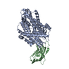
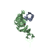
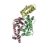
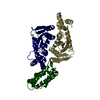
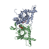
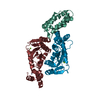
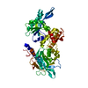
- PDB-1jma: CRYSTAL STRUCTURE OF THE HERPES SIMPLEX VIRUS GLYCOPROTEIN D BOUN... -
+ Open data

- Basic information
| Entry | Database: PDB / ID: 1jma | |||||||||
|---|---|---|---|---|---|---|---|---|---|---|
| Title | CRYSTAL STRUCTURE OF THE HERPES SIMPLEX VIRUS GLYCOPROTEIN D BOUND TO THE CELLULAR RECEPTOR HVEA/HVEM | |||||||||
 Components Components |
| |||||||||
 Keywords Keywords | VIRAL PROTEIN / V-type Ig molecule and TNFR superfamily | |||||||||
| Function / homology |  Function and homology information Function and homology informationnegative regulation of adaptive immune memory response / negative regulation of alpha-beta T cell proliferation / tumor necrosis factor receptor activity / TNFs bind their physiological receptors / positive regulation of cytokine production involved in immune response / Co-inhibition by BTLA / cytokine binding / positive regulation of T cell migration / negative regulation of T cell proliferation / T cell costimulation ...negative regulation of adaptive immune memory response / negative regulation of alpha-beta T cell proliferation / tumor necrosis factor receptor activity / TNFs bind their physiological receptors / positive regulation of cytokine production involved in immune response / Co-inhibition by BTLA / cytokine binding / positive regulation of T cell migration / negative regulation of T cell proliferation / T cell costimulation / virus receptor activity / defense response to Gram-negative bacterium / adaptive immune response / host cell Golgi apparatus / entry receptor-mediated virion attachment to host cell / cell surface receptor signaling pathway / defense response to Gram-positive bacterium / immune response / receptor ligand activity / external side of plasma membrane / innate immune response / viral envelope / symbiont entry into host cell / ubiquitin protein ligase binding / virion membrane / membrane / metal ion binding / plasma membrane Similarity search - Function | |||||||||
| Biological species |   Human herpesvirus 1 (Herpes simplex virus type 1) Human herpesvirus 1 (Herpes simplex virus type 1) Homo sapiens (human) Homo sapiens (human) | |||||||||
| Method |  X-RAY DIFFRACTION / SYNCHROTRON / MIRAS / Resolution: 2.65 Å X-RAY DIFFRACTION / SYNCHROTRON / MIRAS / Resolution: 2.65 Å | |||||||||
 Authors Authors | Carfi, A. / Willis, S.H. / Whitbeck, J.C. / Krummenacker, C. / Cohen, G.H. / Eisenberg, R.J. / Wiley, D.C. | |||||||||
 Citation Citation | Journal: Mol.Cell / Year: 2001 Title: Herpes simplex virus glycoprotein D bound to the human receptor HveA. Authors: Carfi, A. / Willis, S.H. / Whitbeck, J.C. / Krummenacher, C. / Cohen, G.H. / Eisenberg, R.J. / Wiley, D.C. #1: Journal: Cell(Cambridge,Mass.) / Year: 1996Title: Herpes simplex virus-1 entry into cells mediated by a novel member of the TNF/NGF receptor family Authors: Montgomery, R.I. / Warner, M.S. / Lurn, B.J. / Spear, P.G. | |||||||||
| History |
| |||||||||
| Remark 11 | Residues 1 to 3, 93,94 and 106 to 167 including 5 C-terminus HIS tag residues in chain B, and ...Residues 1 to 3, 93,94 and 106 to 167 including 5 C-terminus HIS tag residues in chain B, and residues 260 to 290 that includes 5 C-terimus HIS tag residues were not visible in the electron density maps due to disorder | |||||||||
| Remark 12 | Weak or no electron density was observed for the side chains of the following residues: Chain B ...Weak or no electron density was observed for the side chains of the following residues: Chain B residues: 5, 6, 18, 31, 71, 95 and 96. Chain A residues: 16, 18, 20, 21, 35, 89, 91, 186, 196 and 259 |
- Structure visualization
| Structure viewer | Molecule: MolmilJmol/JSmol |
|---|
- Downloads & links
-Download
| PDBx/mmCIF format | 1jma.cif.gz | 88.4 KB | Display | PDBx/mmCIF format |
|---|---|---|---|---|
| PDB format | pdb1jma.ent.gz | 65.3 KB | Display | PDB format |
| PDBx/mmJSON format | 1jma.json.gz | Tree view | PDBx/mmJSON format | |
| Others |  Other downloads Other downloads |
-Validation report
| Arichive directory | https://data.pdbj.org/pub/pdb/validation_reports/jm/1jmaftp://data.pdbj.org/pub/pdb/validation_reports/jm/1jma | HTTPS FTP |
|---|
-Related structure data


-Links
 PDBj
PDBj
- Assembly
| Deposited unit | 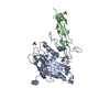
| ||||||||
|---|---|---|---|---|---|---|---|---|---|
| 1 |
| ||||||||
| Unit cell |
|
-Components
| #1: Protein | Mass: 17767.955 Da / Num. of mol.: 1 / Fragment: HVEA-162 / Mutation: ECTODOMAIN OF THE FULL PROTEIN Source method: isolated from a genetically manipulated source Source: (gene. exp.) Human herpesvirus 1 (Herpes simplex virus type 1)Genus: Simplexvirus / Production host:   Spodoptera frugiperda (fall armyworm) / References: UniProt: Q92956 Spodoptera frugiperda (fall armyworm) / References: UniProt: Q92956 | ||||
|---|---|---|---|---|---|
| #2: Protein | Mass: 32315.697 Da / Num. of mol.: 1 / Fragment: GD-285 / Mutation: C-TERMINAL TRUNCATION AT RESIDUE 285 Source method: isolated from a genetically manipulated source Source: (gene. exp.) Homo sapiens (human) / Production host: Spodoptera frugiperda (fall armyworm) / References: UniProt: P57083 | ||||
| #3: Polysaccharide | 2-acetamido-2-deoxy-beta-D-glucopyranose-(1-4)-2-acetamido-2-deoxy-beta-D-glucopyranose Source method: isolated from a genetically manipulated source | ||||
| #4: Chemical | ChemComp-SO4 /   Mass: 96.063 Da / Num. of mol.: 5 / Source method: obtained synthetically / Formula: SO4 Mass: 96.063 Da / Num. of mol.: 5 / Source method: obtained synthetically / Formula: SO4#5: Water | ChemComp-HOH / |  Mass: 18.015 Da / Num. of mol.: 49 / Source method: isolated from a natural source / Formula: H2O Mass: 18.015 Da / Num. of mol.: 49 / Source method: isolated from a natural source / Formula: H2OHas protein modification | Y | |
-Experimental details
-Experiment
| Experiment | Method: X-RAY DIFFRACTION / Number of used crystals: 1 |
|---|
- Sample preparation
| Crystal | Density Matthews: 3.89 Å3/Da / Density % sol: 68.36 % | ||||||||||||||||||||
|---|---|---|---|---|---|---|---|---|---|---|---|---|---|---|---|---|---|---|---|---|---|
| Crystal grow | Temperature: 291 K / Method: vapor diffusion, sitting drop / pH: 8 Details: 1.8M Ammonium Sulfate and 5% Peg400 and Tris-HCL 100mM pH 8.0, VAPOR DIFFUSION, SITTING DROP, temperature 291K | ||||||||||||||||||||
| Crystal grow | *PLUS pH: 8 / Method: unknown | ||||||||||||||||||||
| Components of the solutions | *PLUS
|
-Data collection
| Diffraction | Mean temperature: 100 K |
|---|---|
| Diffraction source | Source: SYNCHROTRON / Site: CHESS  / Beamline: A1 / Wavelength: 0.9196 Å / Beamline: A1 / Wavelength: 0.9196 Å |
| Detector | Type: ADSC QUANTUM 4 / Detector: CCD / Date: May 19, 2000 / Details: monochromator |
| Radiation | Monochromator: GRAPHITE / Protocol: SINGLE WAVELENGTH / Monochromatic (M) / Laue (L): M / Scattering type: x-ray |
| Radiation wavelength | Wavelength: 0.9196 Å / Relative weight: 1 |
| Reflection | Resolution: 2.65→25 Å / Num. all: 24279 / Num. obs: 24260 / % possible obs: 99.7 % / Observed criterion σ(F): 2 / Observed criterion σ(I): 2 / Redundancy: 5 % / Biso Wilson estimate: 57 Å2 / Rmerge(I) obs: 0.068 / Net I/σ(I): 11 |
| Reflection shell | Resolution: 2.65→20 Å / Rmerge(I) obs: 0.346 / Mean I/σ(I) obs: 5 / % possible all: 99.7 |
| Reflection | *PLUS Lowest resolution: 25 Å / Num. measured all: 272205 |
| Reflection shell | *PLUS % possible obs: 99.9 % |
- Processing
| Software |
| |||||||||||||||||||||||||
|---|---|---|---|---|---|---|---|---|---|---|---|---|---|---|---|---|---|---|---|---|---|---|---|---|---|---|
| Refinement | Method to determine structure: MIRAS / Resolution: 2.65→20 Å / σ(F): 1 / Stereochemistry target values: Engh & Huber / Details: mlf refinement with CNS
| |||||||||||||||||||||||||
| Displacement parameters | Biso mean: 16.177 Å2
| |||||||||||||||||||||||||
| Refine analyze | Luzzati sigma a obs: 0.35 Å | |||||||||||||||||||||||||
| Refinement step | Cycle: LAST / Resolution: 2.65→20 Å
| |||||||||||||||||||||||||
| Refine LS restraints |
| |||||||||||||||||||||||||
| Software | *PLUS Name: CNS / Version: 1 / Classification: refinement | |||||||||||||||||||||||||
| Refinement | *PLUS Lowest resolution: 20 Å / σ(F): 1 / % reflection Rfree: 5 % / Rfactor obs: 0.236 | |||||||||||||||||||||||||
| Solvent computation | *PLUS | |||||||||||||||||||||||||
| Displacement parameters | *PLUS | |||||||||||||||||||||||||
| Refine LS restraints | *PLUS Type: c_bond_d / Dev ideal: 0.008 |
