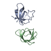
A: SH3-DOMAIN KINASE BINDING PROTEIN 1 B: SH3-DOMAIN KINASE BINDING PROTEIN 1 C: SIGNAL TRANSDUCTION PROTEIN CBL-B SH3-BINDING PROTEIN CBL-B, RING FINGER PROTEIN 56, CBL-B hetero molecules
SH3-DOMAINKINASEBINDINGPROTEIN1 / CBL-INTERACTING PROTEIN OF 85KDA / HUMAN SRC-FAMILY KINASE BINDING PROTEIN 1 / HSB-1 / CD2 BINDING ...CBL-INTERACTING PROTEIN OF 85KDA / HUMAN SRC-FAMILY KINASE BINDING PROTEIN 1 / HSB-1 / CD2 BINDING PROTEIN 3 / CD2BP3 / CIN85
Mass: 6772.526 Da / Num. of mol.: 2 / Fragment: N-TERMINAL SH3 DOMAIN RESIDUES 1-58 Source method: isolated from a genetically manipulated source Source: (gene. exp.) HOMO SAPIENS (human) Organ: BRAIN, KIDNEY, HEART, PLACENTA, LUNG, LIVER, SKELETAL MUSCLE, PANCREAS Plasmid: PET29B / Production host: ESCHERICHIA COLI (E. coli) / Strain (production host): ROSETTA(DE3)PLYS / References: UniProt: Q96B97
Mass: 18.015 Da / Num. of mol.: 82 / Source method: isolated from a natural source / Formula: H2O
-
Experimental details
-
Experiment
Experiment
Method: X-RAY DIFFRACTION / Number of used crystals: 1
-
Sample preparation
Crystal
Density Matthews: 2.4 Å3/Da / Density % sol: 47.3 % Description: STARTING MODEL FOR MOLECULAR REPLACEMENT WAS THE ISOLATED CIN85 N-TERMINAL SH3, UNPUBLISHED.
Crystal grow
pH: 7.5 Details: 0.8 M NA CITRATE, 0.1 M BIS-TRIS PH 7.5, 0.2 M NACL
Type: BRUKER / Detector: CCD / Date: Nov 3, 2004 / Details: MIRRORS
Radiation
Protocol: SINGLE WAVELENGTH / Monochromatic (M) / Laue (L): M / Scattering type: x-ray
Radiation wavelength
Wavelength: 1.5418 Å / Relative weight: 1
Reflection
Resolution: 2→2 Å / Num. obs: 9738 / % possible obs: 99.7 % / Observed criterion σ(I): 0 / Redundancy: 11.8 % / Biso Wilson estimate: 19.1 Å2 / Rmerge(I) obs: 0.06 / Net I/σ(I): 29
Reflection shell
Resolution: 2→2.07 Å / Mean I/σ(I) obs: 2.87 / % possible all: 98.9
-
Processing
Software
Name
Version
Classification
CNS
1.1
refinement
DENZO
datareduction
SCALEPACK
datascaling
AMoRE
phasing
Refinement
Method to determine structure: MOLECULAR REPLACEMENT / Resolution: 2→20 Å / Rfactor Rfree error: 0.012 / Data cutoff high absF: 1029899.76 / Isotropic thermal model: RESTRAINED / Cross valid method: THROUGHOUT / σ(F): 0 / Stereochemistry target values: MAXIMUM LIKELIHOOD Details: A TWINING FRACTION OF 0.305 WAS CONSIDERED DURING REFINEMENT.
In the structure databanks used in Yorodumi, some data are registered as the other names, "COVID-19 virus" and "2019-nCoV". Here are the details of the virus and the list of structure data.
Jan 31, 2019. EMDB accession codes are about to change! (news from PDBe EMDB page)
EMDB accession codes are about to change! (news from PDBe EMDB page)
The allocation of 4 digits for EMDB accession codes will soon come to an end. Whilst these codes will remain in use, new EMDB accession codes will include an additional digit and will expand incrementally as the available range of codes is exhausted. The current 4-digit format prefixed with “EMD-” (i.e. EMD-XXXX) will advance to a 5-digit format (i.e. EMD-XXXXX), and so on. It is currently estimated that the 4-digit codes will be depleted around Spring 2019, at which point the 5-digit format will come into force.
The EM Navigator/Yorodumi systems omit the EMD- prefix.
Related info.:Q: What is EMD? / ID/Accession-code notation in Yorodumi/EM Navigator
Yorodumi is a browser for structure data from EMDB, PDB, SASBDB, etc.
This page is also the successor to EM Navigator detail page, and also detail information page/front-end page for Omokage search.
The word "yorodu" (or yorozu) is an old Japanese word meaning "ten thousand". "mi" (miru) is to see.
Related info.:EMDB / PDB / SASBDB / Comparison of 3 databanks / Yorodumi Search / Aug 31, 2016. New EM Navigator & Yorodumi / Yorodumi Papers / Jmol/JSmol / Function and homology information / Changes in new EM Navigator and Yorodumi
 Movie
Movie Controller


 Open data
 Basic information
 Components
 Keywords
 Function and homology information
 HOMO SAPIENS (human)
 X-RAY DIFFRACTION /
  Authors
 Citation
 Structure visualization
 Downloads & links
 Other downloads
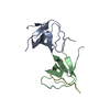


 PDBj
PDBj


 Assembly


 Mass: 22.990 Da / Num. of mol.: 2 / Source method: obtained synthetically / Formula: Na
 Mass: 18.015 Da / Num. of mol.: 82 / Source method: isolated from a natural source / Formula: H2O
 Sample preparation
 / Beamline: 14.2 / Type: BRUKER / Wavelength: 1.5418
  Processing