 Movie
Movie Controller

[English] 日本語
 Yorodumi
- PDB-2bnw: Structural basis for cooperative binding of Ribbon-Helix-Helix Om... -
+ Open data

- Basic information
| Entry | Database: PDB / ID: 2bnw | ||||||
|---|---|---|---|---|---|---|---|
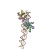
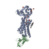
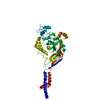
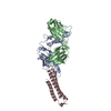
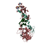
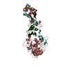
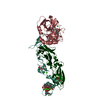
| Title | Structural basis for cooperative binding of Ribbon-Helix-Helix Omega repressor to direct DNA heptad repeats | ||||||
 Components Components |
| ||||||
 Keywords Keywords | DNA-BINDING/REGULATORY PROTEIN / DNA-BINDING-REGULATORY PROTEIN COMPLEX / RIBBON-HELIX-HELIX / RHH / METJ/ARC SUPERFAMILY / COOPERATIVE DNA BINDING / INVERTED REPEATS / DNA HEPTAD / INC18 FAMILY / DNA-BINDING REGULATORY PROTEIN | ||||||
| Function / homology |  Function and homology information Function and homology information | ||||||
| Biological species |  STREPTOCOCCUS PYOGENES (bacteria) STREPTOCOCCUS PYOGENES (bacteria)synthetic construct (others) | ||||||
| Method |  X-RAY DIFFRACTION / SYNCHROTRON / MOLECULAR REPLACEMENT / Resolution: 2.45 Å X-RAY DIFFRACTION / SYNCHROTRON / MOLECULAR REPLACEMENT / Resolution: 2.45 Å | ||||||
 Authors Authors | Weihofen, W.A. / Cicek, A. / Pratto, F. / Alonso, J.C. / Saenger, W. | ||||||
 Citation Citation | Journal: Nucleic Acids Res. / Year: 2006 Title: Structures of Omega Repressors Bound to Direct and Inverted DNA Repeats Explain Modulation of Transcription. Authors: Weihofen, W.A. / Cicek, A. / Pratto, F. / Alonso, J.C. / Saenger, W. | ||||||
| History |
|
- Structure visualization
| Structure viewer | Molecule: MolmilJmol/JSmol |
|---|
- Downloads & links
-Download
| PDBx/mmCIF format | 2bnw.cif.gz | 93.5 KB | Display | PDBx/mmCIF format |
|---|---|---|---|---|
| PDB format | pdb2bnw.ent.gz | 68.2 KB | Display | PDB format |
| PDBx/mmJSON format | 2bnw.json.gz | Tree view | PDBx/mmJSON format | |
| Others |  Other downloads Other downloads |
-Validation report
| Arichive directory | https://data.pdbj.org/pub/pdb/validation_reports/bn/2bnwftp://data.pdbj.org/pub/pdb/validation_reports/bn/2bnw | HTTPS FTP |
|---|
-Related structure data
| Related structure data |  2bnzC  2caxC  1cmaS 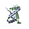 1irqS S: Starting model for refinement C: citing same article ( |
|---|---|
| Similar structure data |

-Links
 PDBj
PDBj


- Assembly
| Deposited unit | 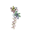
| ||||||||||||||||
|---|---|---|---|---|---|---|---|---|---|---|---|---|---|---|---|---|---|
| 1 |
| ||||||||||||||||
| Unit cell |
| ||||||||||||||||
| Noncrystallographic symmetry (NCS) | NCS oper:
| ||||||||||||||||
| Details | THE DESIGNATION OF THE QUATERNARY STRUCTURE AS OCTAMERICREFLECTS THE STANDARD PQS CONVENTION FOR DESCRIBINGHETEROGENEOUS ASSEMBLIES. HOWEVER, THE CRYSTALLOGRAPHICASYMMETRIC UNIT ACTUALLY CONTAINS ONE DNA FRAGMENT(COMPRISED OF CHAINS E AND F) WHICH IS BOUND TO TWOPROTEIN DIMERS (CHAINS A, B, C AND D). A FURTHER FREEDNA FRAGMENT (CHAINS G AND H) IS PRESENT IN THE A.U.THE INTERFACE BETWEEN THE TWO PROTEIN DIMERS AND DNAIS 1600 ANGSTROMS**2 AND THE INTERFACE BETWEEN THETWO PROTEIN DIMERS IS 280 ANSGTROMS**2. |
-Components
| #1: Protein | Mass: 6137.223 Da / Num. of mol.: 4 / Fragment: RIBBON-HELIX-HELIX DOMAIN, RESIDUES 20-71 Source method: isolated from a genetically manipulated source Source: (gene. exp.) STREPTOCOCCUS PYOGENES (bacteria)Description: OMEGA TRANSCRIPTIONAL REPRESSOR IS ENCODED BY PLASMID PSM19035 OF THE INC18 FAMILY OF PLASMIDS Plasmid: PET28A-DELTA19OMEGA / Production host: #2: DNA chain | Mass: 5486.610 Da / Num. of mol.: 2 / Source method: obtained synthetically Details: DIRECT DNA HEPTAD REPEATS OCCUR IN PROMOTERS PRECEEDING GENES CONTROLLED BY OMEGA TRANSCRIPTIONAL EPRESSOR, INC18 FAMILY OF PLASMIDS Source: (synth.) synthetic construct #3: DNA chain | Mass: 5543.584 Da / Num. of mol.: 2 / Source method: obtained synthetically Details: DIRECT DNA HEPTAD REPEATS OCCUR IN PROMOTERS PRECEEDING GENES CONTROLLED BY OMEGA TRANSCRIPTIONAL EPRESSOR, INC18 FAMILY OF PLASMIDS Source: (synth.) synthetic construct #4: Water | ChemComp-HOH / |  Mass: 18.015 Da / Num. of mol.: 79 / Source method: isolated from a natural source / Formula: H2O Mass: 18.015 Da / Num. of mol.: 79 / Source method: isolated from a natural source / Formula: H2OSequence details | 19 N-TERMINAL RESIDUES TRUNCATED, NEW N-TERMINAL MET19 IS A CLONING ARTEFACT. | |
|---|
-Experimental details
-Experiment
| Experiment | Method: X-RAY DIFFRACTION / Number of used crystals: 1 |
|---|
- Sample preparation
| Crystal | Density Matthews: 4 Å3/Da / Density % sol: 69 % |
|---|---|
| Crystal grow | pH: 7 Details: 150 MM NA/KPO4, PH 7.0, 2.4 NA2MALONATE, PH 7.5, 2% AMINOCAPROIC ACID |
-Data collection
| Diffraction | Mean temperature: 100 K |
|---|---|
| Diffraction source | Source: SYNCHROTRON / Site: BESSY  / Beamline: 14.1 / Wavelength: 1.08 / Beamline: 14.1 / Wavelength: 1.08 |
| Detector | Type: MARRESEARCH / Detector: CCD / Date: Oct 28, 2004 |
| Radiation | Protocol: SINGLE WAVELENGTH / Monochromatic (M) / Laue (L): M / Scattering type: x-ray |
| Radiation wavelength | Wavelength: 1.08 Å / Relative weight: 1 |
| Reflection | Resolution: 2.45→30 Å / Num. obs: 83105 / % possible obs: 97.4 % / Observed criterion σ(I): 1 / Redundancy: 3.3 % / Rmerge(I) obs: 0.06 / Net I/σ(I): 12.7 |
| Reflection shell | Resolution: 2.45→2.54 Å / Redundancy: 2.4 % / Rmerge(I) obs: 0.38 / Mean I/σ(I) obs: 3 / % possible all: 81.7 |
- Processing
| Software |
| ||||||||||||||||||||||||||||||||||||||||||||||||||||||||||||||||||||||||||||||||||||||||||||||||||||||||||||||||||||||||||||||||||||||||||||||||||||||||||||||||||||||||||||||||||||||
|---|---|---|---|---|---|---|---|---|---|---|---|---|---|---|---|---|---|---|---|---|---|---|---|---|---|---|---|---|---|---|---|---|---|---|---|---|---|---|---|---|---|---|---|---|---|---|---|---|---|---|---|---|---|---|---|---|---|---|---|---|---|---|---|---|---|---|---|---|---|---|---|---|---|---|---|---|---|---|---|---|---|---|---|---|---|---|---|---|---|---|---|---|---|---|---|---|---|---|---|---|---|---|---|---|---|---|---|---|---|---|---|---|---|---|---|---|---|---|---|---|---|---|---|---|---|---|---|---|---|---|---|---|---|---|---|---|---|---|---|---|---|---|---|---|---|---|---|---|---|---|---|---|---|---|---|---|---|---|---|---|---|---|---|---|---|---|---|---|---|---|---|---|---|---|---|---|---|---|---|---|---|---|---|
| Refinement | Method to determine structure: MOLECULAR REPLACEMENT Starting model: PDB ENTRY 1IRQ AND 1CMA Resolution: 2.45→43.44 Å / Cor.coef. Fo:Fc: 0.927 / Cor.coef. Fo:Fc free: 0.905 / SU B: 14.188 / SU ML: 0.173 / TLS residual ADP flag: LIKELY RESIDUAL / Cross valid method: THROUGHOUT / ESU R: 0.296 / ESU R Free: 0.236 / Stereochemistry target values: MAXIMUM LIKELIHOOD Details: HYDROGENS HAVE BEEN ADDED IN THE RIDING POSITIONS. CYTOSINES E18 AND G18 WERE ONLY MODELED FOR THE 5'- PHOSPATE AND ATOM C5'
| ||||||||||||||||||||||||||||||||||||||||||||||||||||||||||||||||||||||||||||||||||||||||||||||||||||||||||||||||||||||||||||||||||||||||||||||||||||||||||||||||||||||||||||||||||||||
| Solvent computation | Ion probe radii: 0.8 Å / Shrinkage radii: 0.8 Å / VDW probe radii: 1.2 Å / Solvent model: BABINET MODEL WITH MASK | ||||||||||||||||||||||||||||||||||||||||||||||||||||||||||||||||||||||||||||||||||||||||||||||||||||||||||||||||||||||||||||||||||||||||||||||||||||||||||||||||||||||||||||||||||||||
| Displacement parameters | Biso mean: 45.81 Å2
| ||||||||||||||||||||||||||||||||||||||||||||||||||||||||||||||||||||||||||||||||||||||||||||||||||||||||||||||||||||||||||||||||||||||||||||||||||||||||||||||||||||||||||||||||||||||
| Refinement step | Cycle: LAST / Resolution: 2.45→43.44 Å
| ||||||||||||||||||||||||||||||||||||||||||||||||||||||||||||||||||||||||||||||||||||||||||||||||||||||||||||||||||||||||||||||||||||||||||||||||||||||||||||||||||||||||||||||||||||||
| Refine LS restraints |
|