 Movie
Movie Controller

[English] 日本語
 Yorodumi
- PDB-1irq: Crystal structure of omega transcriptional repressor at 1.5A reso... -
+ Open data

- Basic information
| Entry | Database: PDB / ID: 1irq | ||||||
|---|---|---|---|---|---|---|---|
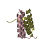
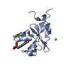
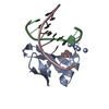
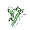
| Title | Crystal structure of omega transcriptional repressor at 1.5A resolution | ||||||
 Components Components | omega transcriptional repressor | ||||||
 Keywords Keywords | GENE REGULATION / transcriptional repressor / ribbon-helix-helix | ||||||
| Function / homology |  Function and homology information Function and homology information | ||||||
| Biological species |  Streptococcus pyogenes (bacteria) Streptococcus pyogenes (bacteria) | ||||||
| Method |  X-RAY DIFFRACTION / SYNCHROTRON / MIR / Resolution: 1.5 Å X-RAY DIFFRACTION / SYNCHROTRON / MIR / Resolution: 1.5 Å | ||||||
 Authors Authors | Murayama, K. / Orth, P. / De La Hoz, A.B. / Alonso, J.C. / Saenger, W. | ||||||
 Citation Citation | Journal: J.Mol.Biol. / Year: 2001 Title: Crystal structure of omega transcriptional repressor encoded by Streptococcus pyogenes plasmid pSM19035 at 1.5 A resolution. Authors: Murayama, K. / Orth, P. / de la Hoz, A.B. / Alonso, J.C. / Saenger, W. | ||||||
| History |
|
- Structure visualization
| Structure viewer | Molecule: MolmilJmol/JSmol |
|---|
- Downloads & links
-Download
| PDBx/mmCIF format | 1irq.cif.gz | 34.2 KB | Display | PDBx/mmCIF format |
|---|---|---|---|---|
| PDB format | pdb1irq.ent.gz | 24.1 KB | Display | PDB format |
| PDBx/mmJSON format | 1irq.json.gz | Tree view | PDBx/mmJSON format | |
| Others |  Other downloads Other downloads |
-Validation report
| Arichive directory | https://data.pdbj.org/pub/pdb/validation_reports/ir/1irqftp://data.pdbj.org/pub/pdb/validation_reports/ir/1irq | HTTPS FTP |
|---|
-Related structure data
| Similar structure data |
|---|


-Links
 PDBj
PDBj- Assembly
| Deposited unit | 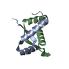
| ||||||||
|---|---|---|---|---|---|---|---|---|---|
| 1 |
| ||||||||
| Unit cell |
|
-Components
| #1: Protein | Mass: 8004.359 Da / Num. of mol.: 2 Source method: isolated from a genetically manipulated source Source: (gene. exp.) Streptococcus pyogenes (bacteria) / Plasmid: pSM19035 / Production host: #2: Water | ChemComp-HOH / |  Mass: 18.015 Da / Num. of mol.: 125 / Source method: isolated from a natural source / Formula: H2O Mass: 18.015 Da / Num. of mol.: 125 / Source method: isolated from a natural source / Formula: H2O |
|---|
-Experimental details
-Experiment
| Experiment | Method: X-RAY DIFFRACTION / Number of used crystals: 1 |
|---|
- Sample preparation
| Crystal | Density Matthews: 1.69 Å3/Da / Density % sol: 27.3 % | ||||||||||||||||||||||||||||||||||||||||||||||||||||||||
|---|---|---|---|---|---|---|---|---|---|---|---|---|---|---|---|---|---|---|---|---|---|---|---|---|---|---|---|---|---|---|---|---|---|---|---|---|---|---|---|---|---|---|---|---|---|---|---|---|---|---|---|---|---|---|---|---|---|
| Crystal grow | Temperature: 291 K / Method: vapor diffusion, hanging drop / pH: 8.5 Details: PEG 3350, 100mM Sodium Acetate, pH 8.5, VAPOR DIFFUSION, HANGING DROP, temperature 291K | ||||||||||||||||||||||||||||||||||||||||||||||||||||||||
| Crystal grow | *PLUS Temperature: 18 ℃ / pH: 7.5 | ||||||||||||||||||||||||||||||||||||||||||||||||||||||||
| Components of the solutions | *PLUS
|
-Data collection
| Diffraction | Mean temperature: 100 K |
|---|---|
| Diffraction source | Source: SYNCHROTRON / Site: EMBL/DESY, HAMBURG  / Beamline: X11 / Wavelength: 0.9073 Å / Beamline: X11 / Wavelength: 0.9073 Å |
| Detector | Type: MARRESEARCH / Detector: CCD |
| Radiation | Protocol: SINGLE WAVELENGTH / Monochromatic (M) / Laue (L): M / Scattering type: x-ray |
| Radiation wavelength | Wavelength: 0.9073 Å / Relative weight: 1 |
| Reflection | Resolution: 1.5→29.6 Å / Num. all: 16703 / Num. obs: 16703 / % possible obs: 97.6 % / Biso Wilson estimate: 24.3 Å2 |
| Reflection | *PLUS Num. measured all: 69483 / Rmerge(I) obs: 0.026 |
| Reflection shell | *PLUS Highest resolution: 1.5 Å / Lowest resolution: 1.52 Å / % possible obs: 95.1 % / Rmerge(I) obs: 0.384 |
- Processing
| Software |
| ||||||||||||||||||||
|---|---|---|---|---|---|---|---|---|---|---|---|---|---|---|---|---|---|---|---|---|---|
| Refinement | Method to determine structure: MIR / Resolution: 1.5→29.6 Å / Rfactor Rfree error: 0.006 / Data cutoff high absF: 652365.39 / Data cutoff low absF: 0 / Isotropic thermal model: RESTRAINED / Cross valid method: THROUGHOUT / σ(F): 3.17
| ||||||||||||||||||||
| Solvent computation | Solvent model: FLAT MODEL / Bsol: 16.6132 Å2 / ksol: 0.317343 e/Å3 | ||||||||||||||||||||
| Displacement parameters | Biso mean: 29.8 Å2
| ||||||||||||||||||||
| Refine analyze |
| ||||||||||||||||||||
| Refinement step | Cycle: LAST / Resolution: 1.5→29.6 Å
| ||||||||||||||||||||
| Refine LS restraints |
| ||||||||||||||||||||
| Xplor file |
| ||||||||||||||||||||
| Software | *PLUS Name: CNS / Version: 1 / Classification: refinement | ||||||||||||||||||||
| Refinement | *PLUS σ(F): 3.17 / % reflection Rfree: 10 % | ||||||||||||||||||||
| Solvent computation | *PLUS | ||||||||||||||||||||
| Displacement parameters | *PLUS Biso mean: 29.8 Å2 | ||||||||||||||||||||
| Refine LS restraints | *PLUS
|
