 Movie
Movie Controller

[English] 日本語
 Yorodumi
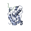
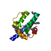
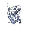
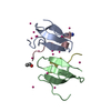
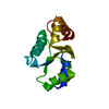
- PDB-1c8c: CRYSTAL STRUCTURES OF THE CHROMOSOMAL PROTEINS SSO7D/SAC7D BOUND ... -
+ Open data

- Basic information
| Entry | Database: PDB / ID: 1c8c | ||||||
|---|---|---|---|---|---|---|---|
| Title | CRYSTAL STRUCTURES OF THE CHROMOSOMAL PROTEINS SSO7D/SAC7D BOUND TO DNA CONTAINING T-G MISMATCHED BASE PAIRS | ||||||
 Components Components |
| ||||||
 Keywords Keywords | DNA BINDING PROTEIN/DNA / DNA BINDING PROTEIN / PROTEIN-DNA INTERACTION / PROTEIN STABILITY / HYPERTHERMOPHILE / ACHAEABACTERIA / ELECTROSTATICS / MOLECULAR MODELING / T-G MISMATCH / DNA BINDING PROTEIN-DNA COMPLEX | ||||||
| Function / homology |  Function and homology information Function and homology information | ||||||
| Biological species |   Sulfolobus solfataricus (archaea) Sulfolobus solfataricus (archaea) | ||||||
| Method |  X-RAY DIFFRACTION / MOLECULAR REPLACEMENT / Resolution: 1.45 Å X-RAY DIFFRACTION / MOLECULAR REPLACEMENT / Resolution: 1.45 Å | ||||||
 Authors Authors | Su, S. / Gao, Y.-G. / Robinson, H. / Liaw, Y.-C. / Edmondson, S.P. / Shriver, J.W. / Wang, A.H.-J. | ||||||
 Citation Citation | Journal: J.Mol.Biol. / Year: 2000 Title: Crystal structures of the chromosomal proteins Sso7d/Sac7d bound to DNA containing T-G mismatched base-pairs. Authors: Su, S. / Gao, Y.G. / Robinson, H. / Liaw, Y.C. / Edmondson, S.P. / Shriver, J.W. / Wang, A.H. | ||||||
| History |
|
- Structure visualization
| Structure viewer | Molecule: MolmilJmol/JSmol |
|---|
- Downloads & links
-Download
| PDBx/mmCIF format | 1c8c.cif.gz | 50.6 KB | Display | PDBx/mmCIF format |
|---|---|---|---|---|
| PDB format | pdb1c8c.ent.gz | 33.2 KB | Display | PDB format |
| PDBx/mmJSON format | 1c8c.json.gz | Tree view | PDBx/mmJSON format | |
| Others |  Other downloads Other downloads |
-Validation report
| Arichive directory | https://data.pdbj.org/pub/pdb/validation_reports/c8/1c8cftp://data.pdbj.org/pub/pdb/validation_reports/c8/1c8c | HTTPS FTP |
|---|
-Related structure data
| Related structure data |  1ca5C  1ca6C  1azpS S: Starting model for refinement C: citing same article ( |
|---|---|
| Similar structure data |
-Links
 PDBj
PDBj- Assembly
| Deposited unit | 
| ||||||||||
|---|---|---|---|---|---|---|---|---|---|---|---|
| 1 |
| ||||||||||
| Unit cell |
|
-Components
| #1: DNA chain | Mass: 2442.616 Da / Num. of mol.: 2 / Source method: obtained synthetically #2: Protein | | Mass: 7236.563 Da / Num. of mol.: 1 / Source method: isolated from a natural source Details: GERMAN COLLECTION OF MICROORGANISMS (DSM) 1617, #1616 Source: (natural) Sulfolobus solfataricus (archaea) / References: UniProt: P39476#3: Water | ChemComp-HOH / |  Mass: 18.015 Da / Num. of mol.: 157 / Source method: isolated from a natural source / Formula: H2O Mass: 18.015 Da / Num. of mol.: 157 / Source method: isolated from a natural source / Formula: H2O |
|---|
-Experimental details
-Experiment
| Experiment | Method: X-RAY DIFFRACTION / Number of used crystals: 1 |
|---|
- Sample preparation
| Crystal | Density Matthews: 1.8 Å3/Da / Density % sol: 39 % | ||||||||||||||||||||||||||||||
|---|---|---|---|---|---|---|---|---|---|---|---|---|---|---|---|---|---|---|---|---|---|---|---|---|---|---|---|---|---|---|---|
| Crystal grow | Method: vapor diffusion, hanging drop / pH: 6.5 Details: PROTEIN WAS CRYSTALLIZED FROM 1.3MM SSO7D, 1.3MM DUPLEX DNA, 2.5 MM TRIS (PH 6.5), 2.5% PEG 400, EQUILIBRATED AGAINST 15% PEG 400 at pH 6.5, VAPOR DIFFUSION, HANGING DROP | ||||||||||||||||||||||||||||||
| Components of the solutions |
| ||||||||||||||||||||||||||||||
| Crystal grow | *PLUS Method: vapor diffusion, sitting drop | ||||||||||||||||||||||||||||||
| Components of the solutions | *PLUS
|
-Data collection
| Diffraction | Mean temperature: 123 K |
|---|---|
| Diffraction source | Source: ROTATING ANODE / Type: RIGAKU RU200 / Wavelength: 1.5418 |
| Detector | Type: RIGAKU RAXIS IIC / Detector: IMAGE PLATE / Date: Aug 15, 1998 |
| Radiation | Monochromator: GRAPHITE / Protocol: SINGLE WAVELENGTH / Monochromatic (M) / Laue (L): M / Scattering type: x-ray |
| Radiation wavelength | Wavelength: 1.5418 Å / Relative weight: 1 |
| Reflection | Resolution: 1.38→20 Å / Num. obs: 14498 / % possible obs: 89.1 % / Observed criterion σ(I): 1 / Rmerge(I) obs: 0.054 |
| Reflection shell | *PLUS Highest resolution: 1.45 Å / Lowest resolution: 1.52 Å / % possible obs: 63 % |
- Processing
| Software |
| ||||||||||||||||||||
|---|---|---|---|---|---|---|---|---|---|---|---|---|---|---|---|---|---|---|---|---|---|
| Refinement | Method to determine structure: MOLECULAR REPLACEMENT Starting model: PDB ENTRY 1AZP Resolution: 1.45→8 Å / Cross valid method: THROUGHOUT / σ(F): 4
| ||||||||||||||||||||
| Refinement step | Cycle: LAST / Resolution: 1.45→8 Å
| ||||||||||||||||||||
| Software | *PLUS Name: SHELXL-97 / Classification: refinement | ||||||||||||||||||||
| Refinement | *PLUS σ(F): 4 / % reflection Rfree: 5 % / Rfactor Rwork: 0.229 | ||||||||||||||||||||
| Solvent computation | *PLUS | ||||||||||||||||||||
| Displacement parameters | *PLUS | ||||||||||||||||||||
| Refine LS restraints | *PLUS
|
