 Movie
Movie Controller

[English] 日本語
 Yorodumi
- PDB-1azp: HYPERTHERMOPHILE CHROMOSOMAL PROTEIN SAC7D BOUND WITH KINKED DNA ... -
+ Open data

- Basic information
| Entry | Database: PDB / ID: 1azp | ||||||
|---|---|---|---|---|---|---|---|
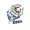
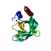
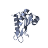
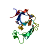
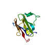
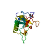
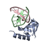
| Title | HYPERTHERMOPHILE CHROMOSOMAL PROTEIN SAC7D BOUND WITH KINKED DNA DUPLEX | ||||||
 Components Components |
| ||||||
 Keywords Keywords | DNA BINDING PROTEIN/DNA / COMPLEX (CHROMATIN PROTEIN-DNA) / DNA-BINDING / ARCHEA KINKED-DNA / MINOR-GROOVE BINDING / INTERCALATION / DNA BINDING PROTEIN-DNA COMPLEX | ||||||
| Function / homology |  Function and homology information Function and homology information | ||||||
| Biological species |   Sulfolobus acidocaldarius (acidophilic) Sulfolobus acidocaldarius (acidophilic) | ||||||
| Method |  X-RAY DIFFRACTION / MIR / Resolution: 1.6 Å X-RAY DIFFRACTION / MIR / Resolution: 1.6 Å | ||||||
 Authors Authors | Robinson, H. / Gao, Y.-G. / Mccrary, B.S. / Edmondson, S.P. / Shriver, J.W. / Wang, A.H.-J. | ||||||
 Citation Citation | Journal: Nature / Year: 1998 Title: The hyperthermophile chromosomal protein Sac7d sharply kinks DNA. Authors: Robinson, H. / Gao, Y.G. / McCrary, B.S. / Edmondson, S.P. / Shriver, J.W. / Wang, A.H. | ||||||
| History |
|
- Structure visualization
| Structure viewer | Molecule: MolmilJmol/JSmol |
|---|
- Downloads & links
-Download
| PDBx/mmCIF format | 1azp.cif.gz | 42 KB | Display | PDBx/mmCIF format |
|---|---|---|---|---|
| PDB format | pdb1azp.ent.gz | 27.1 KB | Display | PDB format |
| PDBx/mmJSON format | 1azp.json.gz | Tree view | PDBx/mmJSON format | |
| Others |  Other downloads Other downloads |
-Validation report
| Arichive directory | https://data.pdbj.org/pub/pdb/validation_reports/az/1azpftp://data.pdbj.org/pub/pdb/validation_reports/az/1azp | HTTPS FTP |
|---|
-Related structure data


-Links
 PDBj
PDBj- Assembly
| Deposited unit | 
| ||||||||||
|---|---|---|---|---|---|---|---|---|---|---|---|
| 1 |
| ||||||||||
| Unit cell |
|
-Components
| #1: DNA chain | Mass: 2427.605 Da / Num. of mol.: 2 / Source method: obtained synthetically #2: Protein | | Mass: 7626.914 Da / Num. of mol.: 1 Source method: isolated from a genetically manipulated source Source: (gene. exp.) Sulfolobus acidocaldarius (acidophilic)Production host:  #3: Water | ChemComp-HOH / |  Mass: 18.015 Da / Num. of mol.: 132 / Source method: isolated from a natural source / Formula: H2O Mass: 18.015 Da / Num. of mol.: 132 / Source method: isolated from a natural source / Formula: H2O |
|---|
-Experimental details
-Experiment
| Experiment | Method: X-RAY DIFFRACTION / Number of used crystals: 1 |
|---|
- Sample preparation
| Crystal | Density Matthews: 2.81 Å3/Da / Density % sol: 56 % | ||||||||||||||||||||||||||||||
|---|---|---|---|---|---|---|---|---|---|---|---|---|---|---|---|---|---|---|---|---|---|---|---|---|---|---|---|---|---|---|---|
| Crystal grow | Method: vapor diffusion, hanging drop / pH: 6.5 / Details: pH 6.5, VAPOR DIFFUSION, HANGING DROP | ||||||||||||||||||||||||||||||
| Components of the solutions |
| ||||||||||||||||||||||||||||||
| Crystal grow | *PLUS | ||||||||||||||||||||||||||||||
| Components of the solutions | *PLUS
|
-Data collection
| Diffraction | Mean temperature: 123 K |
|---|---|
| Diffraction source | Source: ROTATING ANODE / Type: RIGAKU RU200 / Wavelength: 1.540598 |
| Detector | Type: RIGAKU / Detector: IMAGE PLATE / Date: Jul 15, 1997 |
| Radiation | Monochromator: GRAPHITE / Monochromatic (M) / Laue (L): M / Scattering type: x-ray |
| Radiation wavelength | Wavelength: 1.540598 Å / Relative weight: 1 |
| Reflection | Resolution: 1.6→30 Å / Num. obs: 18497 / % possible obs: 95 % / Observed criterion σ(I): 1 / Redundancy: 6.6 % / Biso Wilson estimate: 19.4 Å2 / Rmerge(I) obs: 0.07 / Net I/σ(I): 11.1 |
| Reflection shell | Resolution: 1.6→1.65 Å / Redundancy: 3.7 % / Rmerge(I) obs: 0.393 / Mean I/σ(I) obs: 1.5 / % possible all: 95.2 |
| Reflection shell | *PLUS % possible obs: 95.2 % |
- Processing
| Software |
| ||||||||||||||||||||||||||||||||||||||||||||||||||||||||||||
|---|---|---|---|---|---|---|---|---|---|---|---|---|---|---|---|---|---|---|---|---|---|---|---|---|---|---|---|---|---|---|---|---|---|---|---|---|---|---|---|---|---|---|---|---|---|---|---|---|---|---|---|---|---|---|---|---|---|---|---|---|---|
| Refinement | Method to determine structure: MIR / Resolution: 1.6→6 Å / Rfactor Rfree error: 0.009 / Data cutoff high absF: 100000 / Data cutoff low absF: 0.001 / Cross valid method: A POSTERIORI / σ(F): 3
| ||||||||||||||||||||||||||||||||||||||||||||||||||||||||||||
| Refinement step | Cycle: LAST / Resolution: 1.6→6 Å
| ||||||||||||||||||||||||||||||||||||||||||||||||||||||||||||
| Refine LS restraints |
| ||||||||||||||||||||||||||||||||||||||||||||||||||||||||||||
| Xplor file |
| ||||||||||||||||||||||||||||||||||||||||||||||||||||||||||||
| Software | *PLUS Name: X-PLOR / Version: 3.84 / Classification: refinement | ||||||||||||||||||||||||||||||||||||||||||||||||||||||||||||
| Refinement | *PLUS | ||||||||||||||||||||||||||||||||||||||||||||||||||||||||||||
| Solvent computation | *PLUS | ||||||||||||||||||||||||||||||||||||||||||||||||||||||||||||
| Displacement parameters | *PLUS |
