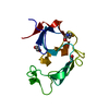
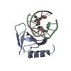
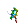
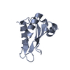
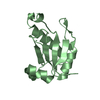
RNA-bindingprotein39 / Coactivator of activating protein 1 and estrogen receptors / Coactivator of AP-1 and ERs / RNA- ...Coactivator of activating protein 1 and estrogen receptors / Coactivator of AP-1 and ERs / RNA-binding motif protein 39 / RNA-binding region-containing protein 2 / Transcription coactivator CAPER
Mass: 12573.253 Da / Num. of mol.: 2 / Fragment: UNP residues 418-530 Source method: isolated from a genetically manipulated source Source: (gene. exp.) Mus musculus (house mouse) / Gene: BC030493, Caper, Rbm39, Rnpc2 / Plasmid: SpeedET / Production host: Escherichia Coli (E. coli) / Strain (production host): HK100 / References: UniProt: Q8VH51
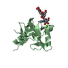
Mass: 18.015 Da / Num. of mol.: 184 / Source method: isolated from a natural source / Formula: H2O
Sequence details
SEQUENCE THE CONSTRUCT WAS EXPRESSED WITH AN N-TERMINAL PURIFICATION TAG MGSDKIHHHHHHENLYFQG. THE ...SEQUENCE THE CONSTRUCT WAS EXPRESSED WITH AN N-TERMINAL PURIFICATION TAG MGSDKIHHHHHHENLYFQG. THE TAG WAS REMOVED WITH TEV PROTEASE LEAVING ONLY A GLYCINE (0) FOLLOWED BY RESIDUES 418-530 OF THE TARGET SEQUENCE. NUMBERING IS BASED ON ISOFORM 1 OF UNIPROTKB Q8VH51.
-
Experimental details
-
Experiment
Experiment
Method: X-RAY DIFFRACTION / Number of used crystals: 1
-
Sample preparation
Crystal
Density Matthews: 2.01 Å3/Da / Density % sol: 38.72 %
Crystal grow
Temperature: 277 K / Method: vapor diffusion, sitting drop / pH: 5 Details: 20.00% polyethylene glycol 6000, 0.1M sodium citrate pH 5.0, NANODROP, VAPOR DIFFUSION, SITTING DROP, temperature 277K
-
Data collection
Diffraction
Mean temperature: 296 K / Ambient temp details: room temperature Crystal support: loop mounted and sealed with a MiTeGen MicroRT polyester capillary
In the structure databanks used in Yorodumi, some data are registered as the other names, "COVID-19 virus" and "2019-nCoV". Here are the details of the virus and the list of structure data.
Jan 31, 2019. EMDB accession codes are about to change! (news from PDBe EMDB page)
EMDB accession codes are about to change! (news from PDBe EMDB page)
The allocation of 4 digits for EMDB accession codes will soon come to an end. Whilst these codes will remain in use, new EMDB accession codes will include an additional digit and will expand incrementally as the available range of codes is exhausted. The current 4-digit format prefixed with “EMD-” (i.e. EMD-XXXX) will advance to a 5-digit format (i.e. EMD-XXXXX), and so on. It is currently estimated that the 4-digit codes will be depleted around Spring 2019, at which point the 5-digit format will come into force.
The EM Navigator/Yorodumi systems omit the EMD- prefix.
Related info.:Q: What is EMD? / ID/Accession-code notation in Yorodumi/EM Navigator
Yorodumi is a browser for structure data from EMDB, PDB, SASBDB, etc.
This page is also the successor to EM Navigator detail page, and also detail information page/front-end page for Omokage search.
The word "yorodu" (or yorozu) is an old Japanese word meaning "ten thousand". "mi" (miru) is to see.
Related info.:EMDB / PDB / SASBDB / Comparison of 3 databanks / Yorodumi Search / Aug 31, 2016. New EM Navigator & Yorodumi / Yorodumi Papers / Jmol/JSmol / Function and homology information / Changes in new EM Navigator and Yorodumi
 Movie
Movie Controller

 Yorodumi
 Open data
 Basic information
 Components
 Keywords
 Function and homology information
 X-RAY DIFFRACTION /
  Authors
 Citation
 Structure visualization
 Downloads & links
 Other downloads
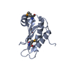


 PDBj
PDBj
 Assembly
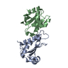

 Mass: 18.015 Da / Num. of mol.: 184 / Source method: isolated from a natural source / Formula: H2O
 Sample preparation
 / Beamline: BL12-2 / Wavelength: 0.9795
  Processing