 Movie
Movie Controller

[English] 日本語
 Yorodumi
- PDB-2bgu: CRYSTAL STRUCTURE OF THE DNA MODIFYING ENZYME BETA-GLUCOSYLTRANSF... -
+ Open data

- Basic information
| Entry | Database: PDB / ID: 2bgu | ||||||
|---|---|---|---|---|---|---|---|
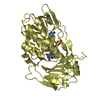
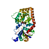
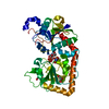
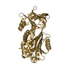
| Title | CRYSTAL STRUCTURE OF THE DNA MODIFYING ENZYME BETA-GLUCOSYLTRANSFERASE IN THE PRESENCE AND ABSENCE OF THE SUBSTRATE URIDINE DIPHOSPHOGLUCOSE | ||||||
 Components Components | BETA-GLUCOSYLTRANSFERASE | ||||||
 Keywords Keywords | TRANSFERASE (GLYCOSYLTRANSFERASE) | ||||||
| Function / homology |  Function and homology information Function and homology informationDNA beta-glucosyltransferase / DNA beta-glucosyltransferase activity / symbiont-mediated evasion of host restriction-modification system / DNA modification / symbiont-mediated suppression of host innate immune response Similarity search - Function | ||||||
| Biological species |  Enterobacteria phage T4 (virus) Enterobacteria phage T4 (virus) | ||||||
| Method |  X-RAY DIFFRACTION / Resolution: 2.2 Å X-RAY DIFFRACTION / Resolution: 2.2 Å | ||||||
 Authors Authors | Vrielink, A. / Rueger, W. / Driessen, H.P.C. / Freemont, P.S. | ||||||
 Citation Citation | Journal: EMBO J. / Year: 1994 Title: Crystal structure of the DNA modifying enzyme beta-glucosyltransferase in the presence and absence of the substrate uridine diphosphoglucose. Authors: Vrielink, A. / Ruger, W. / Driessen, H.P. / Freemont, P.S. #1: Journal: J.Mol.Biol. / Year: 1988Title: Crystallization and Preliminary X-Ray Studies of T4 Phage Beta-Glucosyltransferase Authors: Freemont, P.S. / Ruger, W. #2: Journal: Nucleic Acids Res. / Year: 1985Title: T4-Induced Alpha-and Beta-Glucosyltransferase: Cloning of the Genes and a Comparison of Their Products Based on Sequencing Data Authors: Tomaschewski, J. / Gram, H. / Crabb, J.W. / Ruger, W. | ||||||
| History |
|
- Structure visualization
| Structure viewer | Molecule: MolmilJmol/JSmol |
|---|
- Downloads & links
-Download
| PDBx/mmCIF format | 2bgu.cif.gz | 83.8 KB | Display | PDBx/mmCIF format |
|---|---|---|---|---|
| PDB format | pdb2bgu.ent.gz | 62.4 KB | Display | PDB format |
| PDBx/mmJSON format | 2bgu.json.gz | Tree view | PDBx/mmJSON format | |
| Others |  Other downloads Other downloads |
-Validation report
| Arichive directory | https://data.pdbj.org/pub/pdb/validation_reports/bg/2bguftp://data.pdbj.org/pub/pdb/validation_reports/bg/2bgu | HTTPS FTP |
|---|
-Related structure data

-Links
 PDBj
PDBj


- Assembly
| Deposited unit | 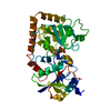
| ||||||||
|---|---|---|---|---|---|---|---|---|---|
| 1 |
| ||||||||
| Unit cell |
|
-Components
| #1: Protein | Mass: 40719.879 Da / Num. of mol.: 1 Source method: isolated from a genetically manipulated source Source: (gene. exp.) Enterobacteria phage T4 (virus) / Genus: T4-like viruses / Species: Enterobacteria phage T4 sensu lato / Production host:  |
|---|---|
| #2: Chemical | ChemComp-UDP /   Type: RNA linking / Mass: 404.161 Da / Num. of mol.: 1 / Source method: obtained synthetically / Formula: C9H14N2O12P2 / Comment: UDP*YM Type: RNA linking / Mass: 404.161 Da / Num. of mol.: 1 / Source method: obtained synthetically / Formula: C9H14N2O12P2 / Comment: UDP*YM |
| #3: Water | ChemComp-HOH /  Mass: 18.015 Da / Num. of mol.: 221 / Source method: isolated from a natural source / Formula: H2O Mass: 18.015 Da / Num. of mol.: 221 / Source method: isolated from a natural source / Formula: H2O |
-Experimental details
-Experiment
| Experiment | Method: X-RAY DIFFRACTION |
|---|
- Sample preparation
| Crystal | Density Matthews: 2.57 Å3/Da / Density % sol: 52.14 % | ||||||||||||||||||||||||||||||||||||||||
|---|---|---|---|---|---|---|---|---|---|---|---|---|---|---|---|---|---|---|---|---|---|---|---|---|---|---|---|---|---|---|---|---|---|---|---|---|---|---|---|---|---|
| Crystal grow | *PLUS Temperature: 18 ℃ / Method: vapor diffusion, hanging drop / PH range low: 5.8 / PH range high: 5.4 | ||||||||||||||||||||||||||||||||||||||||
| Components of the solutions | *PLUS
|
-Data collection
| Reflection | *PLUS Highest resolution: 2.2 Å / Num. obs: 21380 / % possible obs: 94.5 % / Num. measured all: 74613 / Rmerge(I) obs: 0.086 |
|---|
- Processing
| Software |
| ||||||||||||||||||||||||||||||||||||||||||||||||||||||||||||
|---|---|---|---|---|---|---|---|---|---|---|---|---|---|---|---|---|---|---|---|---|---|---|---|---|---|---|---|---|---|---|---|---|---|---|---|---|---|---|---|---|---|---|---|---|---|---|---|---|---|---|---|---|---|---|---|---|---|---|---|---|---|
| Refinement | Resolution: 2.2→10 Å / σ(F): 0 Details: THE COORDINATES ARE PRESENTED IN A COORDINATE FRAME THAT IS TRANSLATED BY 1/4*151.920 ALONG A AND 1/4*52.26 ALONG B. THUS THE TRANSFORMATION PRESENTED ON *SCALE* RECORDS BELOW IS NOT THE ...Details: THE COORDINATES ARE PRESENTED IN A COORDINATE FRAME THAT IS TRANSLATED BY 1/4*151.920 ALONG A AND 1/4*52.26 ALONG B. THUS THE TRANSFORMATION PRESENTED ON *SCALE* RECORDS BELOW IS NOT THE DEFAULT. THE DICTIONARY FOR THE UDP PORTION OF THE SUBSTRATE WAS BUILT USING THE CHARM MINIMIZATION WITHIN QUANTA.
| ||||||||||||||||||||||||||||||||||||||||||||||||||||||||||||
| Refinement step | Cycle: LAST / Resolution: 2.2→10 Å
| ||||||||||||||||||||||||||||||||||||||||||||||||||||||||||||
| Refine LS restraints |
| ||||||||||||||||||||||||||||||||||||||||||||||||||||||||||||
| Software | *PLUS Name: X-PLOR / Classification: refinement | ||||||||||||||||||||||||||||||||||||||||||||||||||||||||||||
| Refinement | *PLUS Rfactor Rwork: 0.191 | ||||||||||||||||||||||||||||||||||||||||||||||||||||||||||||
| Solvent computation | *PLUS | ||||||||||||||||||||||||||||||||||||||||||||||||||||||||||||
| Displacement parameters | *PLUS Biso mean: 21 Å2 |
