 Movie
Movie Controller

[English] 日本語
 Yorodumi
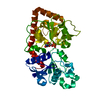
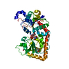
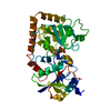
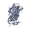
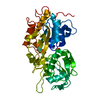
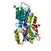
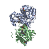
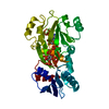
- PDB-1c3j: T4 PHAGE BETA-GLUCOSYLTRANSFERASE: SUBSTRATE BINDING AND PROPOSED... -
+ Open data

- Basic information
| Entry | Database: PDB / ID: 1c3j | ||||||
|---|---|---|---|---|---|---|---|
| Title | T4 PHAGE BETA-GLUCOSYLTRANSFERASE: SUBSTRATE BINDING AND PROPOSED CATALYTIC MECHANISM | ||||||
 Components Components | BETA-GLUCOSYLTRANSFERASE | ||||||
 Keywords Keywords | TRANSFERASE / GLYCOSYLTRANSFERASE | ||||||
| Function / homology |  Function and homology information Function and homology informationDNA beta-glucosyltransferase / DNA beta-glucosyltransferase activity / symbiont-mediated evasion of host restriction-modification system / DNA modification / symbiont-mediated suppression of host innate immune response Similarity search - Function | ||||||
| Biological species |  Enterobacteria phage T4 (virus) Enterobacteria phage T4 (virus) | ||||||
| Method |  X-RAY DIFFRACTION / SYNCHROTRON / Resolution: 1.88 Å X-RAY DIFFRACTION / SYNCHROTRON / Resolution: 1.88 Å | ||||||
 Authors Authors | Morera, S. / Imberty, A. / Aschke-Sonnenborn, U. / Ruger, W. / Freemont, P.S. | ||||||
 Citation Citation | Journal: J.Mol.Biol. / Year: 1999 Title: T4 phage beta-glucosyltransferase: substrate binding and proposed catalytic mechanism. Authors: Morera, S. / Imberty, A. / Aschke-Sonnenborn, U. / Ruger, W. / Freemont, P.S. #1: Journal: Embo J. / Year: 1994Title: Crystal structure of the DNA modifying enzyme beta-glucosyltransferase in the presence and absence of the substrate uridine diphosphoglucose. Authors: Vrielink, A. / Ruger, W. / Driessen, H.P.C. / Freemont, P. | ||||||
| History |
|
- Structure visualization
| Structure viewer | Molecule: MolmilJmol/JSmol |
|---|
- Downloads & links
-Download
| PDBx/mmCIF format | 1c3j.cif.gz | 83.5 KB | Display | PDBx/mmCIF format |
|---|---|---|---|---|
| PDB format | pdb1c3j.ent.gz | 62.4 KB | Display | PDB format |
| PDBx/mmJSON format | 1c3j.json.gz | Tree view | PDBx/mmJSON format | |
| Others |  Other downloads Other downloads |
-Validation report
| Arichive directory | https://data.pdbj.org/pub/pdb/validation_reports/c3/1c3jftp://data.pdbj.org/pub/pdb/validation_reports/c3/1c3j | HTTPS FTP |
|---|
-Related structure data


-Links
 PDBj
PDBj


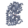
- Assembly
| Deposited unit | 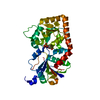
| ||||||||
|---|---|---|---|---|---|---|---|---|---|
| 1 |
| ||||||||
| Unit cell |
|
-Components
| #1: Protein | Mass: 40719.879 Da / Num. of mol.: 1 Source method: isolated from a genetically manipulated source Source: (gene. exp.) Enterobacteria phage T4 (virus) / Genus: T4-like viruses / Species: Enterobacteria phage T4 sensu lato / Production host:  |
|---|---|
| #2: Chemical | ChemComp-UDP /   Type: RNA linking / Mass: 404.161 Da / Num. of mol.: 1 / Source method: obtained synthetically / Formula: C9H14N2O12P2 / Comment: UDP*YM Type: RNA linking / Mass: 404.161 Da / Num. of mol.: 1 / Source method: obtained synthetically / Formula: C9H14N2O12P2 / Comment: UDP*YM |
| #3: Water | ChemComp-HOH /  Mass: 18.015 Da / Num. of mol.: 166 / Source method: isolated from a natural source / Formula: H2O Mass: 18.015 Da / Num. of mol.: 166 / Source method: isolated from a natural source / Formula: H2O |
-Experimental details
-Experiment
| Experiment | Method: X-RAY DIFFRACTION / Number of used crystals: 1 |
|---|
- Sample preparation
| Crystal | Density Matthews: 2.56 Å3/Da / Density % sol: 51.99 % | ||||||||||||||||||||||||||||||
|---|---|---|---|---|---|---|---|---|---|---|---|---|---|---|---|---|---|---|---|---|---|---|---|---|---|---|---|---|---|---|---|
| Crystal grow | Temperature: 291 K / Method: vapor diffusion, hanging drop / pH: 5.6 Details: AMMONIUM SULFATE, pH 5.6, VAPOR DIFFUSION, HANGING DROP, temperature 18K | ||||||||||||||||||||||||||||||
| Crystal grow | *PLUS PH range low: 5.8 / PH range high: 5.4 | ||||||||||||||||||||||||||||||
| Components of the solutions | *PLUS
|
-Data collection
| Diffraction | Mean temperature: 100 K |
|---|---|
| Diffraction source | Source: SYNCHROTRON / Site: LURE  / Beamline: DW32 / Wavelength: 0.97 / Beamline: DW32 / Wavelength: 0.97 |
| Detector | Type: MARRESEARCH / Detector: IMAGE PLATE / Date: Sep 23, 1997 |
| Radiation | Protocol: SINGLE WAVELENGTH / Monochromatic (M) / Laue (L): M / Scattering type: x-ray |
| Radiation wavelength | Wavelength: 0.97 Å / Relative weight: 1 |
| Reflection | Resolution: 1.88→26 Å / Num. all: 223460 / Num. obs: 31888 / % possible obs: 92 % / Observed criterion σ(I): 2 / Redundancy: 6 % / Biso Wilson estimate: 19.659 Å2 / Rmerge(I) obs: 0.05 / Net I/σ(I): 13.5 |
| Reflection shell | Resolution: 1.88→1.98 Å / Redundancy: 3 % / Rmerge(I) obs: 0.278 / % possible all: 71 |
| Reflection | *PLUS Num. measured all: 223460 |
| Reflection shell | *PLUS % possible obs: 71 % |
- Processing
| Software |
| ||||||||||||||||||||||||||||||||||||||||||||||||||||||||||||
|---|---|---|---|---|---|---|---|---|---|---|---|---|---|---|---|---|---|---|---|---|---|---|---|---|---|---|---|---|---|---|---|---|---|---|---|---|---|---|---|---|---|---|---|---|---|---|---|---|---|---|---|---|---|---|---|---|---|---|---|---|---|
| Refinement | Resolution: 1.88→20 Å / σ(F): 2
| ||||||||||||||||||||||||||||||||||||||||||||||||||||||||||||
| Refinement step | Cycle: LAST / Resolution: 1.88→20 Å
| ||||||||||||||||||||||||||||||||||||||||||||||||||||||||||||
| Refine LS restraints |
| ||||||||||||||||||||||||||||||||||||||||||||||||||||||||||||
| Software | *PLUS Name: X-PLOR / Version: 3.843 / Classification: refinement | ||||||||||||||||||||||||||||||||||||||||||||||||||||||||||||
| Refinement | *PLUS | ||||||||||||||||||||||||||||||||||||||||||||||||||||||||||||
| Solvent computation | *PLUS | ||||||||||||||||||||||||||||||||||||||||||||||||||||||||||||
| Displacement parameters | *PLUS Biso mean: 20.67 Å2 |
