 Movie
Movie Controller

[English] 日本語
 Yorodumi
- PDB-1qkj: T4 Phage B-Glucosyltransferase, Substrate Binding and Proposed Ca... -
+ Open data

- Basic information
| Entry | Database: PDB / ID: 1qkj | ||||||
|---|---|---|---|---|---|---|---|
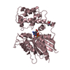
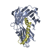
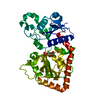
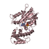
| Title | T4 Phage B-Glucosyltransferase, Substrate Binding and Proposed Catalytic Mechanism | ||||||
 Components Components | BETA-GLUCOSYLTRANSFERASE | ||||||
 Keywords Keywords | TRANSFERASE (GLUCOSYLTRANSFERASE) / TRANSFERASE(GLUCOSYLTRANSFERASE) | ||||||
| Function / homology |  Function and homology information Function and homology informationDNA beta-glucosyltransferase / DNA beta-glucosyltransferase activity / symbiont-mediated evasion of host restriction-modification system / DNA modification / symbiont-mediated suppression of host innate immune response Similarity search - Function | ||||||
| Biological species |  BACTERIOPHAGE T4 (virus) BACTERIOPHAGE T4 (virus) | ||||||
| Method |  X-RAY DIFFRACTION / SYNCHROTRON / MOLECULAR REPLACEMENT / Resolution: 2.3 Å X-RAY DIFFRACTION / SYNCHROTRON / MOLECULAR REPLACEMENT / Resolution: 2.3 Å | ||||||
 Authors Authors | Morera, S. / Imberty, I. / Aschke-Sonnenborn, U. / Ruger, W. / Freemont, P.S. | ||||||
 Citation Citation | Journal: J.Mol.Biol. / Year: 1999 Title: T4 Phage Beta-Glucosyltransferase: Substrate Binding and Proposed Catalytic Mechanism Authors: Morera, S. / Imberty, A. / Aschke-Sonnenborn, U. / Ruger, W. / Freemont, P.S. #1: Journal: Embo J. / Year: 1994Title: Crystal Structure of the DNA Modifying Enzyme Beta-Glucosyltransferase in the Presence and Absence of the Substrate Uridine Diphosphoglucose Authors: Vrielink, A. / Rueger, W. / Driessen, H.P.C. / Freemont, P.S. #2: Journal: J.Mol.Biol. / Year: 1988 Title: Crystallization and Preliminary X-Ray Studies of T4 Phage Beta-Glucosyltransferase Authors: Freemont, P.S. / Rueger, W. #3: Journal: Nucleic Acids Res. / Year: 1985 Title: T4-Induced Alpha- and Beta-Glucosyltransferase: Cloning of the Genes and a Comparison of Their Products Based on Sequencing Data Authors: Tomaschewski, J. / Gram, H. / Crabb, J.W. / Ruger, W. | ||||||
| History |
|
- Structure visualization
| Structure viewer | Molecule: MolmilJmol/JSmol |
|---|
- Downloads & links
-Download
| PDBx/mmCIF format | 1qkj.cif.gz | 87.3 KB | Display | PDBx/mmCIF format |
|---|---|---|---|---|
| PDB format | pdb1qkj.ent.gz | 64.5 KB | Display | PDB format |
| PDBx/mmJSON format | 1qkj.json.gz | Tree view | PDBx/mmJSON format | |
| Others |  Other downloads Other downloads |
-Validation report
| Arichive directory | https://data.pdbj.org/pub/pdb/validation_reports/qk/1qkjftp://data.pdbj.org/pub/pdb/validation_reports/qk/1qkj | HTTPS FTP |
|---|
-Related structure data
| Related structure data |  1c3jC  1bguS S: Starting model for refinement C: citing same article ( |
|---|---|
| Similar structure data |


-Links
 PDBj
PDBj


- Assembly
| Deposited unit | 
| ||||||||
|---|---|---|---|---|---|---|---|---|---|
| 1 |
| ||||||||
| Unit cell |
| ||||||||
| Details | BIOLOGICAL_UNIT: MONOMER |
-Components
| #1: Protein | Mass: 40719.879 Da / Num. of mol.: 1 Source method: isolated from a genetically manipulated source Source: (gene. exp.) BACTERIOPHAGE T4 (virus) / Production host:  |
|---|---|
| #2: Chemical | ChemComp-UDP /   Type: RNA linking / Mass: 404.161 Da / Num. of mol.: 1 / Source method: obtained synthetically / Formula: C9H14N2O12P2 / Comment: UDP*YM Type: RNA linking / Mass: 404.161 Da / Num. of mol.: 1 / Source method: obtained synthetically / Formula: C9H14N2O12P2 / Comment: UDP*YM |
| #3: Water | ChemComp-HOH /  Mass: 18.015 Da / Num. of mol.: 134 / Source method: isolated from a natural source / Formula: H2O Mass: 18.015 Da / Num. of mol.: 134 / Source method: isolated from a natural source / Formula: H2O |
| Compound details | BETA-GLUCOSYLTRANSFERASE TRANSFERS A BETA-D-GLUCOSYL RESIDUE FROM UDP-GLUCOSE TO AN ...BETA-GLUCOSYLTR |
-Experimental details
-Experiment
| Experiment | Method: X-RAY DIFFRACTION / Number of used crystals: 1 |
|---|
- Sample preparation
| Crystal | Density Matthews: 2.6 Å3/Da / Density % sol: 52.63 % | |||||||||||||||||||||||||||||||||||
|---|---|---|---|---|---|---|---|---|---|---|---|---|---|---|---|---|---|---|---|---|---|---|---|---|---|---|---|---|---|---|---|---|---|---|---|---|
| Crystal grow | pH: 7.4 / Details: pH 7.40 | |||||||||||||||||||||||||||||||||||
| Crystal grow | *PLUS pH: 7.5 / Method: vapor diffusion, hanging drop | |||||||||||||||||||||||||||||||||||
| Components of the solutions | *PLUS
|
-Data collection
| Diffraction | Mean temperature: 110 K |
|---|---|
| Diffraction source | Source: SYNCHROTRON / Site: ESRF  / Beamline: BM02 / Wavelength: 1 / Beamline: BM02 / Wavelength: 1 |
| Detector | Detector: CCD |
| Radiation | Protocol: SINGLE WAVELENGTH / Monochromatic (M) / Laue (L): M / Scattering type: x-ray |
| Radiation wavelength | Wavelength: 1 Å / Relative weight: 1 |
| Reflection | Resolution: 2.3→20 Å / Num. obs: 16930 / % possible obs: 96 % / Redundancy: 5 % / Rsym value: 0.41 |
| Reflection | *PLUS Num. measured all: 57848 / Rmerge(I) obs: 0.041 |
| Reflection shell | *PLUS % possible obs: 78 % / Rmerge(I) obs: 0.23 |
- Processing
| Software |
| ||||||||||||||||||||||||||||||||||||||||||||||||||||||||||||
|---|---|---|---|---|---|---|---|---|---|---|---|---|---|---|---|---|---|---|---|---|---|---|---|---|---|---|---|---|---|---|---|---|---|---|---|---|---|---|---|---|---|---|---|---|---|---|---|---|---|---|---|---|---|---|---|---|---|---|---|---|---|
| Refinement | Method to determine structure: MOLECULAR REPLACEMENT Starting model: 1BGU Resolution: 2.3→20 Å / σ(F): 2
| ||||||||||||||||||||||||||||||||||||||||||||||||||||||||||||
| Displacement parameters | Biso mean: 17.49 Å2 | ||||||||||||||||||||||||||||||||||||||||||||||||||||||||||||
| Refinement step | Cycle: LAST / Resolution: 2.3→20 Å
| ||||||||||||||||||||||||||||||||||||||||||||||||||||||||||||
| Refine LS restraints |
| ||||||||||||||||||||||||||||||||||||||||||||||||||||||||||||
| Software | *PLUS Version: 3.84 / Classification: refinement | ||||||||||||||||||||||||||||||||||||||||||||||||||||||||||||
| Refinement | *PLUS Num. reflection obs: 16489 / Rfactor obs: 0.198 / Rfactor Rfree: 0.29 | ||||||||||||||||||||||||||||||||||||||||||||||||||||||||||||
| Solvent computation | *PLUS | ||||||||||||||||||||||||||||||||||||||||||||||||||||||||||||
| Displacement parameters | *PLUS | ||||||||||||||||||||||||||||||||||||||||||||||||||||||||||||
| Refine LS restraints | *PLUS
|
