 Movie
Movie Controller

[English] 日本語
 Yorodumi
- PDB-2apx: Crystal Structure of the G17E/A52V/S54N/K66E/Q72H/E80V/L81S/T87S/... -
+ Open data

- Basic information
| Entry | Database: PDB / ID: 2apx | ||||||
|---|---|---|---|---|---|---|---|
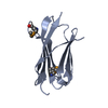
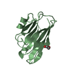
| Title | Crystal Structure of the G17E/A52V/S54N/K66E/Q72H/E80V/L81S/T87S/G96V variant of the murine T cell receptor V beta 8.2 domain | ||||||
 Components Components | T cell receptor beta chain V | ||||||
 Keywords Keywords | IMMUNE SYSTEM / the murine T cell receptor V beta 8.2 domain | ||||||
| Function / homology |  Function and homology information Function and homology information: / Immunoglobulin V-Type / Immunoglobulin V-set domain / Immunoglobulin V-set domain / Ig-like domain profile. / Immunoglobulin-like domain / Immunoglobulin-like domain superfamily / Immunoglobulin-like fold / Immunoglobulins / Immunoglobulin-like ...: / Immunoglobulin V-Type / Immunoglobulin V-set domain / Immunoglobulin V-set domain / Ig-like domain profile. / Immunoglobulin-like domain / Immunoglobulin-like domain superfamily / Immunoglobulin-like fold / Immunoglobulins / Immunoglobulin-like / Sandwich / Mainly Beta Similarity search - Domain/homology | ||||||
| Biological species |   Rattus norvegicus (Norway rat) Rattus norvegicus (Norway rat) | ||||||
| Method |  X-RAY DIFFRACTION / MOLECULAR REPLACEMENT / Resolution: 1.8 Å X-RAY DIFFRACTION / MOLECULAR REPLACEMENT / Resolution: 1.8 Å | ||||||
 Authors Authors | Cho, S. / Swaminathan, C.P. / Yang, J. / Kerzic, M.C. / Guan, R. / Kieke, M.C. / Kranz, D.M. / Mariuzza, R.A. / Sundberg, E.J. | ||||||
 Citation Citation | Journal: Structure / Year: 2005 Title: Structural basis of affinity maturation and intramolecular cooperativity in a protein-protein interaction. Authors: Cho, S. / Swaminathan, C.P. / Yang, J. / Kerzic, M.C. / Guan, R. / Kieke, M.C. / Kranz, D.M. / Mariuzza, R.A. / Sundberg, E.J. | ||||||
| History |
| ||||||
| Remark 999 | SEQUENCE NO SUITABLE SEQUENCE DATABASE REFERENCE WAS AVAILABLE AT THE TIME OF PROCESSING THIS FILE. |
- Structure visualization
| Structure viewer | Molecule: MolmilJmol/JSmol |
|---|
- Downloads & links
-Download
| PDBx/mmCIF format | 2apx.cif.gz | 40.1 KB | Display | PDBx/mmCIF format |
|---|---|---|---|---|
| PDB format | pdb2apx.ent.gz | 27 KB | Display | PDB format |
| PDBx/mmJSON format | 2apx.json.gz | Tree view | PDBx/mmJSON format | |
| Others |  Other downloads Other downloads |
-Validation report
| Arichive directory | https://data.pdbj.org/pub/pdb/validation_reports/ap/2apxftp://data.pdbj.org/pub/pdb/validation_reports/ap/2apx | HTTPS FTP |
|---|
-Related structure data
| Related structure data |  2apbC  2apfC  2aptC  2apvC  2apwC  2aq1C 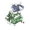 2aq2C  2aq3C C: citing same article ( |
|---|---|
| Similar structure data |




-Links
 PDBj
PDBj
- Assembly
| Deposited unit | 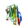
| ||||||||||||
|---|---|---|---|---|---|---|---|---|---|---|---|---|---|
| 1 |
| ||||||||||||
| Unit cell |
| ||||||||||||
| Components on special symmetry positions |
|
-Components
| #1: Protein | Mass: 12184.484 Da / Num. of mol.: 1 Mutation: G17E, A52V, S54N, K66E, Q72H, E80V, L81S, T87S, G96V Source method: isolated from a genetically manipulated source Source: (gene. exp.) Rattus norvegicus (Norway rat) / Plasmid: pT7-7 / Production host:   Escherichia coli (E. coli) / Strain (production host): BL21(DE3)pLysS / References: UniProt: A2NAI0 Escherichia coli (E. coli) / Strain (production host): BL21(DE3)pLysS / References: UniProt: A2NAI0 | ||||
|---|---|---|---|---|---|
| #2: Chemical | 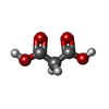  Mass: 104.061 Da / Num. of mol.: 2 / Source method: obtained synthetically / Formula: C3H4O4 Mass: 104.061 Da / Num. of mol.: 2 / Source method: obtained synthetically / Formula: C3H4O4#3: Water | ChemComp-HOH / |  Mass: 18.015 Da / Num. of mol.: 190 / Source method: isolated from a natural source / Formula: H2O Mass: 18.015 Da / Num. of mol.: 190 / Source method: isolated from a natural source / Formula: H2OHas protein modification | Y | |
-Experimental details
-Experiment
| Experiment | Method: X-RAY DIFFRACTION / Number of used crystals: 1 |
|---|
- Sample preparation
| Crystal | Density Matthews: 2.8 Å3/Da / Density % sol: 55.3 % |
|---|---|
| Crystal grow | Temperature: 298 K / Method: vapor diffusion, hanging drop / pH: 7 Details: 2.0 M Sodium Malonate, 0.2 % dioxane, pH 7, VAPOR DIFFUSION, HANGING DROP, temperature 298K |
-Data collection
| Diffraction | Mean temperature: 100 K |
|---|---|
| Diffraction source | Source: ROTATING ANODE / Type: RIGAKU RU200 / Wavelength: 1.5418 Å |
| Detector | Type: RIGAKU RAXIS IV / Detector: IMAGE PLATE / Date: Nov 13, 2003 / Details: mirrors |
| Radiation | Monochromator: OSMIC MIRRORS / Protocol: SINGLE WAVELENGTH / Monochromatic (M) / Laue (L): M / Scattering type: x-ray |
| Radiation wavelength | Wavelength: 1.5418 Å / Relative weight: 1 |
| Reflection | Resolution: 1.44→37.3 Å / Num. all: 25200 / Num. obs: 21554 / % possible obs: 85.5 % / Observed criterion σ(F): 2 / Observed criterion σ(I): 4 / Limit h max: 22 / Limit h min: 0 / Limit k max: 51 / Limit k min: 0 / Limit l max: 78 / Limit l min: 0 |
| Reflection shell | Resolution: 1.44→1.595 Å / % possible all: 37 |
- Processing
| Software |
| ||||||||||||||||||||||||||||||||||||||||||||||||||||||||||||||||||||||||||||||||||||||||||
|---|---|---|---|---|---|---|---|---|---|---|---|---|---|---|---|---|---|---|---|---|---|---|---|---|---|---|---|---|---|---|---|---|---|---|---|---|---|---|---|---|---|---|---|---|---|---|---|---|---|---|---|---|---|---|---|---|---|---|---|---|---|---|---|---|---|---|---|---|---|---|---|---|---|---|---|---|---|---|---|---|---|---|---|---|---|---|---|---|---|---|---|
| Refinement | Method to determine structure: MOLECULAR REPLACEMENT / Resolution: 1.8→30 Å / Cor.coef. Fo:Fc: 0.954 / Cor.coef. Fo:Fc free: 0.935 / SU B: 2.515 / SU ML: 0.079 / Cross valid method: THROUGHOUT / σ(F): 0 / ESU R: 0.123 / ESU R Free: 0.123 / Stereochemistry target values: MAXIMUM LIKELIHOOD
| ||||||||||||||||||||||||||||||||||||||||||||||||||||||||||||||||||||||||||||||||||||||||||
| Solvent computation | Ion probe radii: 0.8 Å / Shrinkage radii: 0.8 Å / VDW probe radii: 1.2 Å / Solvent model: MASK | ||||||||||||||||||||||||||||||||||||||||||||||||||||||||||||||||||||||||||||||||||||||||||
| Displacement parameters | Biso mean: 20.017 Å2
| ||||||||||||||||||||||||||||||||||||||||||||||||||||||||||||||||||||||||||||||||||||||||||
| Refinement step | Cycle: LAST / Resolution: 1.8→30 Å
| ||||||||||||||||||||||||||||||||||||||||||||||||||||||||||||||||||||||||||||||||||||||||||
| Refine LS restraints |
| ||||||||||||||||||||||||||||||||||||||||||||||||||||||||||||||||||||||||||||||||||||||||||
| LS refinement shell | Resolution: 1.8→1.847 Å / Total num. of bins used: 20
|
