Protocol: SINGLE WAVELENGTH / Monochromatic (M) / Laue (L): M / Scattering type: x-ray
Radiation wavelength
Wavelength: 0.9793 Å / Relative weight: 1
Reflection
Resolution: 4.5→20 Å / Num. obs: 4479 / % possible obs: 100 %
Reflection shell
Resolution: 4.5→4.66 Å / % possible all: 100
-
Processing
Software
Name
Version
Classification
NB
REFMAC
5.2.0005
refinement
PDB_EXTRACT
1.7
dataextraction
DENZO
datareduction
SCALEPACK
datascaling
PHASER
phasing
Refinement
Method to determine structure: MOLECULAR REPLACEMENT / Resolution: 4.5→20 Å / Cor.coef. Fo:Fc: 0.91 / Cor.coef. Fo:Fc free: 0.866 / SU B: 180.892 / SU ML: 1.005 / TLS residual ADP flag: LIKELY RESIDUAL / Cross valid method: THROUGHOUT / σ(F): 0 / ESU R Free: 1.3 / Stereochemistry target values: MAXIMUM LIKELIHOOD Details: POORLY DEFINED ELECTRON DENSITY WAS OBSERVED FOR SEVERAL SIDE CHAINS. NEVERTHELESS THEIR INCLUSION LEAD TO BETTER BEHAVIOUR DURING REFINEMENT, ACCORDING TO THE RFREE VALUE, AND THE RFREE/RWORK DIFFERENTIAL
Rfactor
Num. reflection
% reflection
Selection details
Rfree
0.346
321
7.6 %
RANDOM
Rwork
0.271
-
-
-
all
0.276
-
-
-
obs
-
4215
95.82 %
-
Solvent computation
Ion probe radii: 0.8 Å / Shrinkage radii: 1.4 Å / VDW probe radii: 1.4 Å / Solvent model: MASK
Displacement parameters
Biso mean: 166.369 Å2
Baniso -1
Baniso -2
Baniso -3
1-
12.96 Å2
0 Å2
0 Å2
2-
-
12.96 Å2
0 Å2
3-
-
-
-25.93 Å2
Refinement step
Cycle: LAST / Resolution: 4.5→20 Å
Protein
Nucleic acid
Ligand
Solvent
Total
Num. atoms
2468
0
6
0
2474
Refine LS restraints
Refine-ID
Type
Dev ideal
Dev ideal target
Number
X-RAY DIFFRACTION
r_bond_refined_d
0.013
0.022
2535
X-RAY DIFFRACTION
r_angle_refined_deg
1.786
1.967
3453
X-RAY DIFFRACTION
r_dihedral_angle_1_deg
9.621
5
321
X-RAY DIFFRACTION
r_dihedral_angle_2_deg
39.846
25.739
115
X-RAY DIFFRACTION
r_dihedral_angle_3_deg
25.133
15
418
X-RAY DIFFRACTION
r_dihedral_angle_4_deg
14.518
15
9
X-RAY DIFFRACTION
r_chiral_restr
0.142
0.2
397
X-RAY DIFFRACTION
r_gen_planes_refined
0.004
0.02
1936
X-RAY DIFFRACTION
r_nbd_refined
0.297
0.2
1472
X-RAY DIFFRACTION
r_nbtor_refined
0.326
0.2
1675
X-RAY DIFFRACTION
r_xyhbond_nbd_refined
0.233
0.2
100
X-RAY DIFFRACTION
r_metal_ion_refined
0.322
0.2
20
X-RAY DIFFRACTION
r_symmetry_vdw_refined
0.806
0.2
125
X-RAY DIFFRACTION
r_symmetry_hbond_refined
0.538
0.2
8
X-RAY DIFFRACTION
r_mcbond_it
0.245
1.5
1638
X-RAY DIFFRACTION
r_mcangle_it
0.445
2
2613
X-RAY DIFFRACTION
r_scbond_it
0.507
3
987
X-RAY DIFFRACTION
r_scangle_it
0.892
4.5
840
LS refinement shell
Resolution: 4.5→4.736 Å / Total num. of bins used: 10
Rfactor
Num. reflection
% reflection
Rfree
0.39
38
-
Rwork
0.325
505
-
all
-
543
-
obs
-
-
90.35 %
Refinement TLS params.
Method: refined / Refine-ID: X-RAY DIFFRACTION
ID
L11 (°2)
L12 (°2)
L13 (°2)
L22 (°2)
L23 (°2)
L33 (°2)
S11 (Å °)
S12 (Å °)
S13 (Å °)
S21 (Å °)
S22 (Å °)
S23 (Å °)
S31 (Å °)
S32 (Å °)
S33 (Å °)
T11 (Å2)
T12 (Å2)
T13 (Å2)
T22 (Å2)
T23 (Å2)
T33 (Å2)
Origin x (Å)
Origin y (Å)
Origin z (Å)
1
5.863
0.4537
-4.1716
8.3445
0.7209
12.0052
-0.6164
0.1034
-0.0351
0.9899
-0.5532
0.5726
-0.0061
-2.5002
1.1696
-0.4945
0.061
0.0261
0.4646
0.1766
-0.2245
-78.9166
-86.643
261.8446
2
12.0405
1.8652
-12.5968
2.1168
-5.9689
29.5966
-0.04
-0.2095
-0.3454
-0.2632
-0.0098
0.68
-0.0484
-0.1777
0.0498
-0.7159
0.031
0.011
-0.9103
-0.0009
-0.571
-47.0701
-83.7868
229.4645
3
9.2523
-2.9753
-4.864
9.3704
12.8447
55.2841
-0.6448
0.1907
0.6518
-0.3176
0.5473
0.8588
0.3539
0.0765
0.0976
-0.572
0.0228
-0.1093
-0.732
0.2613
-0.418
-33.4782
-83.7254
183.828
Refinement TLS group
Refine-ID: X-RAY DIFFRACTION / Selection: ALL / Auth asym-ID: A / Label asym-ID: A
ID
Refine TLS-ID
Auth seq-ID
Label seq-ID
1
1
1 - 98
1 - 98
2
2
99 - 207
99 - 207
3
3
208 - 322
208 - 322
+
About Yorodumi
-
News
-
Feb 9, 2022. New format data for meta-information of EMDB entries
New format data for meta-information of EMDB entries
Version 3 of the EMDB header file is now the official format.
The previous official version 1.9 will be removed from the archive.
In the structure databanks used in Yorodumi, some data are registered as the other names, "COVID-19 virus" and "2019-nCoV". Here are the details of the virus and the list of structure data.
Jan 31, 2019. EMDB accession codes are about to change! (news from PDBe EMDB page)
EMDB accession codes are about to change! (news from PDBe EMDB page)
The allocation of 4 digits for EMDB accession codes will soon come to an end. Whilst these codes will remain in use, new EMDB accession codes will include an additional digit and will expand incrementally as the available range of codes is exhausted. The current 4-digit format prefixed with “EMD-” (i.e. EMD-XXXX) will advance to a 5-digit format (i.e. EMD-XXXXX), and so on. It is currently estimated that the 4-digit codes will be depleted around Spring 2019, at which point the 5-digit format will come into force.
The EM Navigator/Yorodumi systems omit the EMD- prefix.
Related info.:Q: What is EMD? / ID/Accession-code notation in Yorodumi/EM Navigator
Yorodumi is a browser for structure data from EMDB, PDB, SASBDB, etc.
This page is also the successor to EM Navigator detail page, and also detail information page/front-end page for Omokage search.
The word "yorodu" (or yorozu) is an old Japanese word meaning "ten thousand". "mi" (miru) is to see.
Related info.:EMDB / PDB / SASBDB / Comparison of 3 databanks / Yorodumi Search / Aug 31, 2016. New EM Navigator & Yorodumi / Yorodumi Papers / Jmol/JSmol / Function and homology information / Changes in new EM Navigator and Yorodumi
 Movie
Movie Controller


 Open data
 Basic information
 Components
 Keywords
 Function and homology information
 X-RAY DIFFRACTION /
  Authors
 Citation
 Structure visualization
 Downloads & links
 Other downloads


 PDBj
PDBj
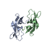
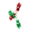
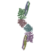
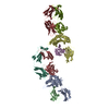
 Assembly


 Mass: 40.078 Da / Num. of mol.: 6 / Source method: obtained synthetically / Formula: Ca
 Sample preparation
 / Beamline: 31-ID / Wavelength: 0.9793 Å
 Processing