- PDB-2a1k: RB69 single-stranded DNA binding protein core domain -
+
Open data
ID or keywords:
Loading...
-
Basic information
Entry
Database: PDB / ID: 2a1k
Title
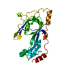
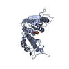
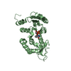
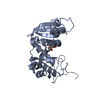
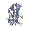
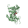
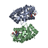
RB69 single-stranded DNA binding protein core domain
Components
gp32 single stranded DNA binding protein
Keywords
DNA BINDING PROTEIN / Zn2+ binding subdomain / 5-stranded beta-sheet / OB fold / single-stranded DNA binding
Function / homology
Function and homology information
bidirectional double-stranded viral DNA replication / single-stranded DNA binding / DNA recombination / DNA replication / DNA repair / metal ion binding Similarity search - Function
Replication Fork Single-Stranded DNA Binding Protein / Replication Fork Single-Stranded Dna Binding Protein / Bacteriophage T4, Gp32, single-stranded DNA-binding domain / Bacteriophage T4, Gp32, single-stranded DNA-binding superfamily / Bacteriophage T4, Gp32, single-stranded DNA-binding / gp32 DNA binding protein like / Nucleic acid-binding, OB-fold / Alpha-Beta Complex / Alpha Beta Similarity search - Domain/homology
Journal: Proteins / Year: 2006 Title: Structure and enzymatic properties of a chimeric bacteriophage RB69 DNA polymerase and single-stranded DNA binding protein with increased processivity. Authors: Sun, S. / Geng, L. / Shamoo, Y.
In the structure databanks used in Yorodumi, some data are registered as the other names, "COVID-19 virus" and "2019-nCoV". Here are the details of the virus and the list of structure data.
Jan 31, 2019. EMDB accession codes are about to change! (news from PDBe EMDB page)
EMDB accession codes are about to change! (news from PDBe EMDB page)
The allocation of 4 digits for EMDB accession codes will soon come to an end. Whilst these codes will remain in use, new EMDB accession codes will include an additional digit and will expand incrementally as the available range of codes is exhausted. The current 4-digit format prefixed with “EMD-” (i.e. EMD-XXXX) will advance to a 5-digit format (i.e. EMD-XXXXX), and so on. It is currently estimated that the 4-digit codes will be depleted around Spring 2019, at which point the 5-digit format will come into force.
The EM Navigator/Yorodumi systems omit the EMD- prefix.
Related info.:Q: What is EMD? / ID/Accession-code notation in Yorodumi/EM Navigator
Yorodumi is a browser for structure data from EMDB, PDB, SASBDB, etc.
This page is also the successor to EM Navigator detail page, and also detail information page/front-end page for Omokage search.
The word "yorodu" (or yorozu) is an old Japanese word meaning "ten thousand". "mi" (miru) is to see.
Related info.:EMDB / PDB / SASBDB / Comparison of 3 databanks / Yorodumi Search / Aug 31, 2016. New EM Navigator & Yorodumi / Yorodumi Papers / Jmol/JSmol / Function and homology information / Changes in new EM Navigator and Yorodumi
 Movie
Movie Controller


 Open data
 Basic information
 Components
 Keywords
 Function and homology information
 Enterobacteria phage RB69 (virus)
 X-RAY DIFFRACTION /
  Authors
 Citation
 Structure visualization
 Downloads & links
 Other downloads
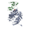


 PDBj
PDBj Assembly


 Mass: 65.409 Da / Num. of mol.: 2 / Source method: obtained synthetically / Formula: Zn
 Mass: 18.015 Da / Num. of mol.: 436 / Source method: isolated from a natural source / Formula: H2O
 Sample preparation
 Processing