- PDB-2atq: RB69 single-stranded DNA binding protein-DNA polymerase fusion -
+
Open data
ID or keywords:
Loading...
-
Basic information
Entry
Database: PDB / ID: 2atq
Title
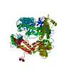
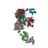
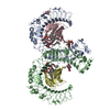
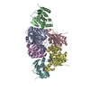
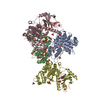
RB69 single-stranded DNA binding protein-DNA polymerase fusion
Components
DNA polymerase
gp32
Keywords
TRANSFERASE/DNA BINDING PROTEIN / DNA polymerase / palm domain / fingers domain / thumb domain / single-stranded DNA binding protein / OB-fold / TRANSFERASE-DNA BINDING PROTEIN COMPLEX
Function / homology
Function and homology information
bidirectional double-stranded viral DNA replication / Hydrolases; Acting on ester bonds; Exodeoxyribonucleases producing 5'-phosphomonoesters / 3'-5' exonuclease activity / DNA-templated DNA replication / single-stranded DNA binding / DNA recombination / DNA-directed DNA polymerase / DNA-directed DNA polymerase activity / DNA replication / nucleotide binding ...bidirectional double-stranded viral DNA replication / Hydrolases; Acting on ester bonds; Exodeoxyribonucleases producing 5'-phosphomonoesters / 3'-5' exonuclease activity / DNA-templated DNA replication / single-stranded DNA binding / DNA recombination / DNA-directed DNA polymerase / DNA-directed DNA polymerase activity / DNA replication / nucleotide binding / DNA repair / DNA binding / metal ion binding Similarity search - Function
Replication Fork Single-Stranded DNA Binding Protein / Replication Fork Single-Stranded Dna Binding Protein / Bacteriophage T4, Gp32, single-stranded DNA-binding domain / Bacteriophage T4, Gp32, single-stranded DNA-binding superfamily / Bacteriophage T4, Gp32, single-stranded DNA-binding / gp32 DNA binding protein like / Monooxygenase - #300 / DNA-directed DNA polymerase T4 type / DNA Polymerase; Chain A, domain 1 / DNA Polymerase, chain B, domain 1 ...Replication Fork Single-Stranded DNA Binding Protein / Replication Fork Single-Stranded Dna Binding Protein / Bacteriophage T4, Gp32, single-stranded DNA-binding domain / Bacteriophage T4, Gp32, single-stranded DNA-binding superfamily / Bacteriophage T4, Gp32, single-stranded DNA-binding / gp32 DNA binding protein like / Monooxygenase - #300 / DNA-directed DNA polymerase T4 type / DNA Polymerase; Chain A, domain 1 / DNA Polymerase, chain B, domain 1 / B family DNA polymerase, finger domain / Palm domain of DNA polymerase / B family DNA polymerase, palm domain / Monooxygenase / : / DNA-directed DNA polymerase, family B, multifunctional domain / DNA-directed DNA polymerase, family B, conserved site / DNA polymerase family B signature. / DNA polymerase family B / DNA polymerase family B, exonuclease domain / DNA-directed DNA polymerase, family B, exonuclease domain / DNA polymerase, palm domain superfamily / DNA polymerase type-B family / DNA-directed DNA polymerase, family B / Ribonuclease H-like superfamily/Ribonuclease H / Nucleotidyltransferase; domain 5 / Helix Hairpins / Ribonuclease H superfamily / Ribonuclease H-like superfamily / Nucleic acid-binding, OB-fold / DNA/RNA polymerase superfamily / Alpha-Beta Complex / Up-down Bundle / 2-Layer Sandwich / Orthogonal Bundle / Mainly Alpha / Alpha Beta Similarity search - Domain/homology
GUANOSINE-5'-DIPHOSPHATE / DNA-directed DNA polymerase / Single-stranded DNA-binding protein Similarity search - Component
Biological species
Enterobacteria phage RB69 (virus)
Method
X-RAY DIFFRACTION / SYNCHROTRON / MAD / Resolution: 3.2 Å
Journal: Proteins / Year: 2006 Title: Structure and enzymatic properties of a chimeric bacteriophage RB69 DNA polymerase and single-stranded DNA binding protein with increased processivity. Authors: Sun, S. / Geng, L. / Shamoo, Y.
SEQUENCE THE C-TERMINUS OF RB69 SINGLE-STRANDED DNA BINDING PROTEIN CORE DOMAIN IS FUSED TO THE N- ...SEQUENCE THE C-TERMINUS OF RB69 SINGLE-STRANDED DNA BINDING PROTEIN CORE DOMAIN IS FUSED TO THE N-TERMINUS OF DNA POLYMERASE THROUGH A LINKER CONSISTING OF GTGSGT. THE LINKER WAS NOT OBSERVED IN THE DENSITY.
Mass: 104655.141 Da / Num. of mol.: 1 / Mutation: D222A, D327A Source method: isolated from a genetically manipulated source Details: C-terminus is fused to a linker not seen in the density Source: (gene. exp.) Enterobacteria phage RB69 (virus) / Genus: T4-like viruses / Gene: 43 / Plasmid: pET101 / Species (production host): Escherichia coli / Production host: Escherichia coli BL21(DE3) (bacteria) / Strain (production host): BL21(DE3) / References: UniProt: Q38087, DNA-directed DNA polymerase
#2: Protein
gp32
Mass: 26228.523 Da / Num. of mol.: 1 / Fragment: RB69 single-stranded DNA binding protein Source method: isolated from a genetically manipulated source Details: N-terminus is fused to a linker not seen in the density Source: (gene. exp.) Enterobacteria phage RB69 (virus) / Genus: T4-like viruses / Gene: gp32 / Plasmid: pET101 / Species (production host): Escherichia coli / Production host: Escherichia coli BL21(DE3) (bacteria) / Strain (production host): BL21(DE3) / References: UniProt: Q7Y265
In the structure databanks used in Yorodumi, some data are registered as the other names, "COVID-19 virus" and "2019-nCoV". Here are the details of the virus and the list of structure data.
Jan 31, 2019. EMDB accession codes are about to change! (news from PDBe EMDB page)
EMDB accession codes are about to change! (news from PDBe EMDB page)
The allocation of 4 digits for EMDB accession codes will soon come to an end. Whilst these codes will remain in use, new EMDB accession codes will include an additional digit and will expand incrementally as the available range of codes is exhausted. The current 4-digit format prefixed with “EMD-” (i.e. EMD-XXXX) will advance to a 5-digit format (i.e. EMD-XXXXX), and so on. It is currently estimated that the 4-digit codes will be depleted around Spring 2019, at which point the 5-digit format will come into force.
The EM Navigator/Yorodumi systems omit the EMD- prefix.
Related info.:Q: What is EMD? / ID/Accession-code notation in Yorodumi/EM Navigator
Yorodumi is a browser for structure data from EMDB, PDB, SASBDB, etc.
This page is also the successor to EM Navigator detail page, and also detail information page/front-end page for Omokage search.
The word "yorodu" (or yorozu) is an old Japanese word meaning "ten thousand". "mi" (miru) is to see.
Related info.:EMDB / PDB / SASBDB / Comparison of 3 databanks / Yorodumi Search / Aug 31, 2016. New EM Navigator & Yorodumi / Yorodumi Papers / Jmol/JSmol / Function and homology information / Changes in new EM Navigator and Yorodumi
 Movie
Movie Controller


 Open data
 Basic information
 Components
 Keywords
 Function and homology information
 Enterobacteria phage RB69 (virus)
 X-RAY DIFFRACTION /
  Authors
 Citation
 Structure visualization
 Downloads & links
 Other downloads
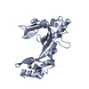


 PDBj
PDBj


 Assembly
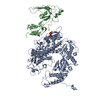


 Type: RNA linking / Mass: 443.201 Da / Num. of mol.: 1 / Source method: obtained synthetically / Formula: C10H15N5O11P2 / Comment: GDP, energy-carrying molecule*YM
 Mass: 65.409 Da / Num. of mol.: 1 / Source method: obtained synthetically / Formula: Zn
 Sample preparation
 / Beamline: 19-ID / Wavelength: 0.95372 Å
 Processing