 Movie
Movie Controller


+ Open data

- Basic information
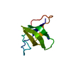
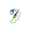
| Entry | Database: PDB / ID: 1zue | ||||||
|---|---|---|---|---|---|---|---|
| Title | Revised Solution Structure of DLP-2 | ||||||
 Components Components | Defensin-like peptide 2/4 | ||||||
 Keywords Keywords | TOXIN / helix / antiparallel beta-sheet | ||||||
| Function / homology |  Function and homology information Function and homology information | ||||||
| Method | SOLUTION NMR / distance geometry, simulated annealing, molecular dynamics, torsion angle dynamics | ||||||
 Authors Authors | Torres, A.M. / Tsampazi, C. / Geraghty, D.P. / Bansal, P.S. / Alewood, P.F. / Kuchel, P.W. | ||||||
 Citation Citation | Journal: Biochem.J. / Year: 2005 Title: D-amino acid residue in a defensin-like peptide from platypus venom: effect on structure and chromatographic properties. Authors: Torres, A.M. / Tsampazi, C. / Geraghty, D.P. / Bansal, P.S. / Alewood, P.F. / Kuchel, P.W. #1: Journal: Biochem.J. / Year: 2000Title: Defensin-like peptide-2 from platypus venom: member of a class of peptides with a distinct structural fold Authors: Torres, A.M. / de Plater, G.M. / Doverskog, M. / Birinyi-Strachan, L.C. / Nicholson, G.M. / Gallagher, C.H. / Kuchel, P.W. | ||||||
| History |
|
- Structure visualization
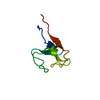
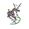
| Structure viewer | Molecule: MolmilJmol/JSmol |
|---|
- Downloads & links
-Download
| PDBx/mmCIF format | 1zue.cif.gz | 268.4 KB | Display | PDBx/mmCIF format |
|---|---|---|---|---|
| PDB format | pdb1zue.ent.gz | 222.1 KB | Display | PDB format |
| PDBx/mmJSON format | 1zue.json.gz | Tree view | PDBx/mmJSON format | |
| Others |  Other downloads Other downloads |
-Validation report
| Arichive directory | https://data.pdbj.org/pub/pdb/validation_reports/zu/1zueftp://data.pdbj.org/pub/pdb/validation_reports/zu/1zue | HTTPS FTP |
|---|
-Related structure data


-Links
 PDBj
PDBj- Assembly
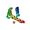
| Deposited unit | 
| |||||||||
|---|---|---|---|---|---|---|---|---|---|---|
| 1 |
| |||||||||
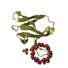
| NMR ensembles |
|
-Components
| #1: Protein/peptide | Mass: 5121.899 Da / Num. of mol.: 1 / Source method: obtained synthetically / Details: chemically synthesized / References: UniProt: P82140 |
|---|---|
| Has protein modification | Y |
-Experimental details
-Experiment
| Experiment | Method: SOLUTION NMR |
|---|---|
| NMR experiment | Type: 2D NOESY |
| NMR details | Text: This structure was determined using standard 2D homonuclear techniques |
- Sample preparation
| Details | Contents: 1mM DLP-2 / Solvent system: 90% H20, 10% D2O |
|---|---|
| Sample conditions | pH: 3.6 / Pressure: ambient / Temperature: 298 K |
-NMR measurement
| NMR spectrometer | Type: Bruker AVANCE / Manufacturer: Bruker / Model: AVANCE / Field strength: 600 MHz |
|---|
- Processing
| NMR software |
| ||||||||||||||||||||||||||||||||
|---|---|---|---|---|---|---|---|---|---|---|---|---|---|---|---|---|---|---|---|---|---|---|---|---|---|---|---|---|---|---|---|---|---|
| Refinement | Method: distance geometry, simulated annealing, molecular dynamics, torsion angle dynamics Software ordinal: 1 Details: The structures are based on a total of 739 restraints, 699 are NOE-derived distance constraints, 24 dihedral angle restraints, 16 distance restraints from hydrogen bonds. | ||||||||||||||||||||||||||||||||
| NMR representative | Selection criteria: closest to the average | ||||||||||||||||||||||||||||||||
| NMR ensemble | Conformer selection criteria: structures with the lowest energy Conformers calculated total number: 500 / Conformers submitted total number: 20 |
