 Movie
Movie Controller

[English] 日本語
 Yorodumi
- PDB-1zow: Crystal Structure of S. aureus FabH, beta-ketoacyl carrier protei... -
+ Open data

- Basic information
| Entry | Database: PDB / ID: 1zow | ||||||
|---|---|---|---|---|---|---|---|
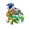
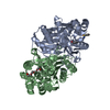
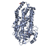
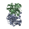
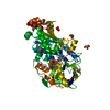
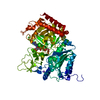
| Title | Crystal Structure of S. aureus FabH, beta-ketoacyl carrier protein synthase III | ||||||
 Components Components | 3-oxoacyl-[acyl-carrier-protein] synthase III | ||||||
 Keywords Keywords | TRANSFERASE / FabH / fatty acid biosynthesis | ||||||
| Function / homology |  Function and homology information Function and homology informationbeta-ketoacyl-[acyl-carrier-protein] synthase III / beta-ketoacyl-acyl-carrier-protein synthase III activity / 3-oxoacyl-[acyl-carrier-protein] synthase activity / fatty acid biosynthetic process / cytoplasm Similarity search - Function | ||||||
| Biological species |   Staphylococcus aureus subsp. aureus (bacteria) Staphylococcus aureus subsp. aureus (bacteria) | ||||||
| Method |  X-RAY DIFFRACTION / SYNCHROTRON / MOLECULAR REPLACEMENT / Resolution: 2 Å X-RAY DIFFRACTION / SYNCHROTRON / MOLECULAR REPLACEMENT / Resolution: 2 Å | ||||||
 Authors Authors | Qiu, X. / Choudhry, A.E. / Janson, C.A. / Grooms, M. / Daines, R.A. / Lonsdale, J.T. / Khandekar, S.S. | ||||||
 Citation Citation | Journal: Protein Sci. / Year: 2005 Title: Crystal structure and substrate specificity of the beta-ketoacyl-acyl carrier protein synthase III (FabH) from Staphylococcus aureus. Authors: Qiu, X. / Choudhry, A.E. / Janson, C.A. / Grooms, M. / Daines, R.A. / Lonsdale, J.T. / Khandekar, S.S. | ||||||
| History |
| ||||||
| Remark 999 | SEQUENCE According to the author residues VAL60, GLU171 and LEU287 are correct based on the electron density. |
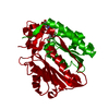
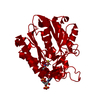
- Structure visualization
| Structure viewer | Molecule: MolmilJmol/JSmol |
|---|
- Downloads & links
-Download
| PDBx/mmCIF format | 1zow.cif.gz | 254.6 KB | Display | PDBx/mmCIF format |
|---|---|---|---|---|
| PDB format | pdb1zow.ent.gz | 206.4 KB | Display | PDB format |
| PDBx/mmJSON format | 1zow.json.gz | Tree view | PDBx/mmJSON format | |
| Others |  Other downloads Other downloads |
-Validation report
| Arichive directory | https://data.pdbj.org/pub/pdb/validation_reports/zo/1zowftp://data.pdbj.org/pub/pdb/validation_reports/zo/1zow | HTTPS FTP |
|---|
-Related structure data
| Related structure data | 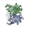 1hn9S S: Starting model for refinement |
|---|---|
| Similar structure data |

-Links
 PDBj
PDBj

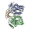
- Assembly
| Deposited unit | 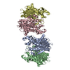
| ||||||||
|---|---|---|---|---|---|---|---|---|---|
| 1 | 
| ||||||||
| 2 | 
| ||||||||
| Unit cell |
|
-Components
| #1: Protein | Mass: 33916.398 Da / Num. of mol.: 4 Source method: isolated from a genetically manipulated source Source: (gene. exp.) Staphylococcus aureus subsp. aureus (bacteria)Species: Staphylococcus aureus / Strain: MW2 / Production host: References: UniProt: Q8NXE2, beta-ketoacyl-[acyl-carrier-protein] synthase I #2: Water | ChemComp-HOH / |  Mass: 18.015 Da / Num. of mol.: 792 / Source method: isolated from a natural source / Formula: H2O Mass: 18.015 Da / Num. of mol.: 792 / Source method: isolated from a natural source / Formula: H2O |
|---|
-Experimental details
-Experiment
| Experiment | Method: X-RAY DIFFRACTION / Number of used crystals: 1 |
|---|
- Sample preparation
| Crystal | Density Matthews: 2.2 Å3/Da / Density % sol: 50 % |
|---|---|
| Crystal grow | Temperature: 273 K / Method: vapor diffusion, sitting drop / pH: 5.6 Details: PEG3K, pH 5.6, VAPOR DIFFUSION, SITTING DROP, temperature 273K |
-Data collection
| Diffraction | Mean temperature: 100 K |
|---|---|
| Diffraction source | Source: SYNCHROTRON / Site: APS  / Beamline: 17-ID / Wavelength: 1 Å / Beamline: 17-ID / Wavelength: 1 Å |
| Detector | Type: ADSC QUANTUM 4 / Detector: CCD / Date: Feb 10, 2001 |
| Radiation | Protocol: SINGLE WAVELENGTH / Monochromatic (M) / Laue (L): M / Scattering type: x-ray |
| Radiation wavelength | Wavelength: 1 Å / Relative weight: 1 |
| Reflection | Resolution: 2→20 Å / Num. obs: 84128 / % possible obs: 96.6 % / Observed criterion σ(F): 2 / Observed criterion σ(I): 2 / Rmerge(I) obs: 0.085 |
| Reflection shell | Resolution: 2→2.07 Å / Rmerge(I) obs: 0.231 / Mean I/σ(I) obs: 2.7 / % possible all: 99.9 |
- Processing
| Software |
| ||||||||||||||||||||
|---|---|---|---|---|---|---|---|---|---|---|---|---|---|---|---|---|---|---|---|---|---|
| Refinement | Method to determine structure: MOLECULAR REPLACEMENT Starting model: PDB entry 1HN9 Resolution: 2→20 Å / σ(F): 0 / Stereochemistry target values: Engh & Huber
| ||||||||||||||||||||
| Refinement step | Cycle: LAST / Resolution: 2→20 Å
| ||||||||||||||||||||
| Refine LS restraints |
|
