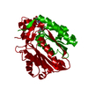
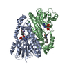
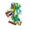
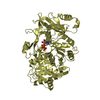
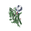
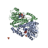
symbiotic process benefiting host / xyloglucan catabolic process / xyloglucan-specific endo-beta-1,4-glucanase / xyloglucan-specific endo-beta-1,4-glucanase activity / cellulase activity / cell outer membrane Similarity search - Function
Glycosyl hydrolases family 9 (GH9) active site signature 1. / Cellulase N-terminal ig-like domain / Cellulase, Ig-like domain / Glycoside hydrolase family 9, His active site / Glycosyl hydrolases family 9 (GH9) active site signature 2. / Glycosyl hydrolases family 9, Asp/Glu active sites / Glycosyl hydrolases family 9 (GH9) active site signature 3. / Glycoside hydrolase family 9 / Glycosyl hydrolase family 9 / Six-hairpin glycosidase-like superfamily ...Glycosyl hydrolases family 9 (GH9) active site signature 1. / Cellulase N-terminal ig-like domain / Cellulase, Ig-like domain / Glycoside hydrolase family 9, His active site / Glycosyl hydrolases family 9 (GH9) active site signature 2. / Glycosyl hydrolases family 9, Asp/Glu active sites / Glycosyl hydrolases family 9 (GH9) active site signature 3. / Glycoside hydrolase family 9 / Glycosyl hydrolase family 9 / Six-hairpin glycosidase-like superfamily / Six-hairpin glycosidase superfamily / Prokaryotic membrane lipoprotein lipid attachment site profile. / Immunoglobulin E-set / Immunoglobulin-like fold Similarity search - Domain/homology
In the structure databanks used in Yorodumi, some data are registered as the other names, "COVID-19 virus" and "2019-nCoV". Here are the details of the virus and the list of structure data.
Jan 31, 2019. EMDB accession codes are about to change! (news from PDBe EMDB page)
EMDB accession codes are about to change! (news from PDBe EMDB page)
The allocation of 4 digits for EMDB accession codes will soon come to an end. Whilst these codes will remain in use, new EMDB accession codes will include an additional digit and will expand incrementally as the available range of codes is exhausted. The current 4-digit format prefixed with “EMD-” (i.e. EMD-XXXX) will advance to a 5-digit format (i.e. EMD-XXXXX), and so on. It is currently estimated that the 4-digit codes will be depleted around Spring 2019, at which point the 5-digit format will come into force.
The EM Navigator/Yorodumi systems omit the EMD- prefix.
Related info.:Q: What is EMD? / ID/Accession-code notation in Yorodumi/EM Navigator
Yorodumi is a browser for structure data from EMDB, PDB, SASBDB, etc.
This page is also the successor to EM Navigator detail page, and also detail information page/front-end page for Omokage search.
The word "yorodu" (or yorozu) is an old Japanese word meaning "ten thousand". "mi" (miru) is to see.
Related info.:EMDB / PDB / SASBDB / Comparison of 3 databanks / Yorodumi Search / Aug 31, 2016. New EM Navigator & Yorodumi / Yorodumi Papers / Jmol/JSmol / Function and homology information / Changes in new EM Navigator and Yorodumi
 Movie
Movie Controller


 Open data
 Basic information
 Components
 Keywords
 Function and homology information
 Bacteroides ovatus (bacteria)
 X-RAY DIFFRACTION /
  Authors
 United States, 1items
  Citation
 Structure visualization
 Downloads & links
 Other downloads
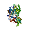


 PDBj
PDBj


 Assembly
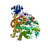


 Mass: 69.085 Da / Num. of mol.: 1 / Source method: obtained synthetically / Formula: C3H5N2
 Mass: 40.078 Da / Num. of mol.: 2 / Source method: obtained synthetically / Formula: Ca
 Mass: 62.068 Da / Num. of mol.: 14 / Source method: obtained synthetically / Formula: C2H6O2
 Mass: 106.120 Da / Num. of mol.: 3 / Source method: obtained synthetically / Formula: C4H10O3
 Mass: 35.453 Da / Num. of mol.: 2 / Source method: obtained synthetically / Formula: Cl
 Mass: 24.305 Da / Num. of mol.: 1 / Source method: obtained synthetically / Formula: Mg
 Sample preparation
 Processing