 Movie
Movie Controller

[English] 日本語
 Yorodumi
- PDB-1zo3: The P-site and P/E-site tRNA structures fitted to P/I site codon. -
+ Open data

- Basic information
| Entry | Database: PDB / ID: 1zo3 | ||||||
|---|---|---|---|---|---|---|---|
| Title | The P-site and P/E-site tRNA structures fitted to P/I site codon. | ||||||
 Components Components | tRNA | ||||||
 Keywords Keywords | RNA / E. COLI / RIBOSOME / INITIATION OF PROTEIN SYNTHESIS / INITIATION FACTOR / CRYO-ELETRON MICROSCOPY | ||||||
| Function / homology | RNA / RNA (> 10) Function and homology information Function and homology information | ||||||
| Biological species |  | ||||||
| Method | ELECTRON MICROSCOPY / single particle reconstruction / cryo EM / Resolution: 13.8 Å | ||||||
 Authors Authors | Allen, G.S. / Zavialov, A. / Gursky, R. / Ehrenberg, M. / Frank, J. | ||||||
 Citation Citation | Journal: Cell / Year: 2005 Title: The cryo-EM structure of a translation initiation complex from Escherichia coli. Authors: Gregory S Allen / Andrey Zavialov / Richard Gursky / Måns Ehrenberg / Joachim Frank /  Abstract: The 70S ribosome and its complement of factors required for initiation of translation in E. coli were purified separately and reassembled in vitro with GDPNP, producing a stable initiation complex ...The 70S ribosome and its complement of factors required for initiation of translation in E. coli were purified separately and reassembled in vitro with GDPNP, producing a stable initiation complex (IC) stalled after 70S assembly. We have obtained a cryo-EM reconstruction of the IC showing IF2*GDPNP at the intersubunit cleft of the 70S ribosome. IF2*GDPNP contacts the 30S and 50S subunits as well as fMet-tRNA(fMet). IF2 here adopts a conformation radically different from that seen in the recent crystal structure of IF2. The C-terminal domain of IF2 binds to the single-stranded portion of fMet-tRNA(fMet), thereby forcing the tRNA into a novel orientation at the P site. The GTP binding domain of IF2 binds to the GTPase-associated center of the 50S subunit in a manner similar to EF-G and EF-Tu. Additionally, we present evidence for the localization of IF1, IF3, one C-terminal domain of L7/L12, and the N-terminal domain of IF2 in the initiation complex. | ||||||
| History |
|
- Structure visualization
| Movie |
Movie viewer |
|---|---|
| Structure viewer | Molecule: MolmilJmol/JSmol |
- Downloads & links
-Download
| PDBx/mmCIF format | 1zo3.cif.gz | 104.1 KB | Display | PDBx/mmCIF format |
|---|---|---|---|---|
| PDB format | pdb1zo3.ent.gz | 56 KB | Display | PDB format |
| PDBx/mmJSON format | 1zo3.json.gz | Tree view | PDBx/mmJSON format | |
| Others |  Other downloads Other downloads |
-Validation report
| Arichive directory | https://data.pdbj.org/pub/pdb/validation_reports/zo/1zo3ftp://data.pdbj.org/pub/pdb/validation_reports/zo/1zo3 | HTTPS FTP |
|---|
-Related structure data
| Related structure data | 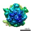 1248MC  1249MC  1zo1C C: citing same article ( M: map data used to model this data |
|---|---|
| Similar structure data |
-Links
 PDBj
PDBj


- Assembly
| Deposited unit | 
|
|---|---|
| 1 |
|
-Components
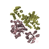
| #1: RNA chain | Mass: 24518.570 Da / Num. of mol.: 2 / Source method: isolated from a natural source / Details: P-site and P/E-site tRNA / Source: (natural) |
|---|
-Experimental details
-Experiment
| Experiment | Method: ELECTRON MICROSCOPY |
|---|---|
| EM experiment | Aggregation state: PARTICLE / 3D reconstruction method: single particle reconstruction |
- Sample preparation
| Component | Name: 70S INITIATION COMPLEX / Type: RIBOSOME |
|---|---|
| Buffer solution | Name: POLYMIX BUFFER / pH: 7.5 / Details: POLYMIX BUFFER |
| Specimen | Conc.: 0.032 mg/ml / Embedding applied: NO / Shadowing applied: NO / Staining applied: NO / Vitrification applied: YES |
| Vitrification | Instrument: FEI VITROBOT MARK I / Cryogen name: ETHANE Details: HOLEY CARBON GRID AT 20C, FLASH FROZEN INTO LIQUID ETHAN |
- Electron microscopy imaging
| Experimental equipment |  Model: Tecnai F20 / Image courtesy: FEI Company |
|---|---|
| Microscopy | Model: FEI TECNAI F20 / Date: Jan 1, 2004 / Details: LOW DOSE MODE |
| Electron gun | Electron source:  FIELD EMISSION GUN / Accelerating voltage: 200 kV / Illumination mode: FLOOD BEAM FIELD EMISSION GUN / Accelerating voltage: 200 kV / Illumination mode: FLOOD BEAM |
| Electron lens | Mode: BRIGHT FIELD / Nominal magnification: 39000 X / Nominal defocus max: -3930 nm / Nominal defocus min: -930 nm / Cs: 2 mm |
| Specimen holder | Temperature: 100 K / Tilt angle max: 0 ° / Tilt angle min: 0 ° |
| Image recording | Film or detector model: KODAK SO-163 FILM |
- Processing
| CTF correction | Details: DEFOCUS GROUPS 0.93-3.93 UM | ||||||||||||
|---|---|---|---|---|---|---|---|---|---|---|---|---|---|
| Symmetry | Point symmetry: C1 (asymmetric) | ||||||||||||
| 3D reconstruction | Method: MULTI-REFERENCE / Resolution: 13.8 Å / Resolution method: FSC 0.5 CUT-OFF / Num. of particles: 20283 / Nominal pixel size: 2.82 Å / Actual pixel size: 2.82 Å Details: resolution 13.8 ANGSTROMS (FSC=0.5) and 8.6 ANGSTROMS (3sigma) Symmetry type: POINT | ||||||||||||
| Atomic model building | Protocol: RIGID BODY FIT / Space: REAL / Target criteria: BEST FIT USING RSREF Details: REFINEMENT PROTOCOL--RIGID BODY FIT OF TRNA DETAILS--the tRNA coordinate files were fitted to the E. coli translation initiation complex map (EMBL-EMD 3525) in the following way- first the ...Details: REFINEMENT PROTOCOL--RIGID BODY FIT OF TRNA DETAILS--the tRNA coordinate files were fitted to the E. coli translation initiation complex map (EMBL-EMD 3525) in the following way- first the cross-correlation of the respective 30S subunit maps were maximized and the resulting transformation matrix was computed. second the coordinates of the tRNA models were transformed according to the matrix previously computed. | ||||||||||||
| Refinement step | Cycle: LAST
|
