 Movie
Movie Controller

[English] 日本語
 Yorodumi
- PDB-1znd: Strong Solute-Solute Dispersive Interactions in a Protein-Ligand ... -
+ Open data

- Basic information
| Entry | Database: PDB / ID: 1znd | ||||||
|---|---|---|---|---|---|---|---|
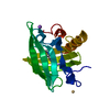
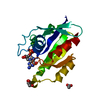
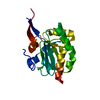
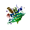
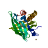
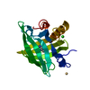
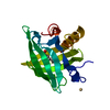
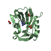
| Title | Strong Solute-Solute Dispersive Interactions in a Protein-Ligand Complex | ||||||
 Components Components | Major Urinary Protein | ||||||
 Keywords Keywords | TRANSPORT PROTEIN / LIPOCALIN / BETA-BARREL | ||||||
| Function / homology |  Function and homology information Function and homology informationpheromone binding / negative regulation of lipid biosynthetic process / positive regulation of glucose metabolic process / odorant binding / energy reserve metabolic process / insulin receptor activity / negative regulation of insulin secretion involved in cellular response to glucose stimulus / heat generation / cellular response to lipid / positive regulation of lipid metabolic process ...pheromone binding / negative regulation of lipid biosynthetic process / positive regulation of glucose metabolic process / odorant binding / energy reserve metabolic process / insulin receptor activity / negative regulation of insulin secretion involved in cellular response to glucose stimulus / heat generation / cellular response to lipid / positive regulation of lipid metabolic process / locomotor rhythm / small molecule binding / negative regulation of lipid storage / negative regulation of gluconeogenesis / aerobic respiration / mitochondrion organization / glucose homeostasis / positive regulation of phosphatidylinositol 3-kinase/protein kinase B signal transduction / negative regulation of DNA-templated transcription / positive regulation of gene expression / : / nucleus / cytosol Similarity search - Function | ||||||
| Biological species |  | ||||||
| Method |  X-RAY DIFFRACTION / MOLECULAR REPLACEMENT / Resolution: 1.6 Å X-RAY DIFFRACTION / MOLECULAR REPLACEMENT / Resolution: 1.6 Å | ||||||
 Authors Authors | Malham, R. / Johnstone, S. / Bingham, R.J. / Barratt, E. / Phillips, S.E. / Laughton, C.A. / Homans, S.W. | ||||||
 Citation Citation | Journal: J.Am.Chem.Soc. / Year: 2005 Title: Strong Solute-Solute Dispersive Interactions in a Protein-Ligand Complex. Authors: Malham, R. / Johnstone, S. / Bingham, R.J. / Barratt, E. / Phillips, S.E. / Laughton, C.A. / Homans, S.W. | ||||||
| History |
|
- Structure visualization
| Structure viewer | Molecule: MolmilJmol/JSmol |
|---|
- Downloads & links
-Download
| PDBx/mmCIF format | 1znd.cif.gz | 51.4 KB | Display | PDBx/mmCIF format |
|---|---|---|---|---|
| PDB format | pdb1znd.ent.gz | 35.7 KB | Display | PDB format |
| PDBx/mmJSON format | 1znd.json.gz | Tree view | PDBx/mmJSON format | |
| Others |  Other downloads Other downloads |
-Validation report
| Arichive directory | https://data.pdbj.org/pub/pdb/validation_reports/zn/1zndftp://data.pdbj.org/pub/pdb/validation_reports/zn/1znd | HTTPS FTP |
|---|
-Related structure data
| Related structure data |  1zneC 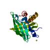 1zngC 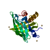 1znhC  1znkC  1znlC  1qy0S C: citing same article ( S: Starting model for refinement |
|---|---|
| Similar structure data |


-Links
 PDBj
PDBj

- Assembly
| Deposited unit | 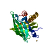
| ||||||||
|---|---|---|---|---|---|---|---|---|---|
| 1 |
| ||||||||
| Unit cell |
|
-Components
| #1: Protein | Mass: 20139.400 Da / Num. of mol.: 1 Source method: isolated from a genetically manipulated source Source: (gene. exp.)  | ||||||
|---|---|---|---|---|---|---|---|
| #2: Chemical |   Mass: 112.411 Da / Num. of mol.: 2 / Source method: obtained synthetically / Formula: Cd Mass: 112.411 Da / Num. of mol.: 2 / Source method: obtained synthetically / Formula: Cd#3: Chemical |   Mass: 88.148 Da / Num. of mol.: 2 / Source method: obtained synthetically / Formula: C5H12O Mass: 88.148 Da / Num. of mol.: 2 / Source method: obtained synthetically / Formula: C5H12O#4: Water | ChemComp-HOH / |  Mass: 18.015 Da / Num. of mol.: 206 / Source method: isolated from a natural source / Formula: H2O Mass: 18.015 Da / Num. of mol.: 206 / Source method: isolated from a natural source / Formula: H2OHas protein modification | Y | |
-Experimental details
-Experiment
| Experiment | Method: X-RAY DIFFRACTION / Number of used crystals: 1 |
|---|
- Sample preparation
| Crystal | Density Matthews: 2.72 Å3/Da / Density % sol: 54.44 % |
|---|---|
| Crystal grow | Temperature: 291 K / Method: vapor diffusion, hanging drop / pH: 4.9 Details: CdCl, malate, HCl, pH 4.9, VAPOR DIFFUSION, HANGING DROP, temperature 291K |
-Data collection
| Diffraction | Mean temperature: 100 K |
|---|---|
| Diffraction source | Source: ROTATING ANODE / Type: RIGAKU / Wavelength: 1.5418 Å |
| Detector | Type: RIGAKU RAXIS IV / Detector: IMAGE PLATE / Date: Apr 12, 2005 / Details: confocal max flux (osmic) |
| Radiation | Monochromator: Ni filter / Protocol: SINGLE WAVELENGTH / Monochromatic (M) / Laue (L): M / Scattering type: x-ray |
| Radiation wavelength | Wavelength: 1.5418 Å / Relative weight: 1 |
| Reflection | Resolution: 1.6→19.5 Å / Num. all: 26264 / Num. obs: 26264 / % possible obs: 96.6 % / Observed criterion σ(F): 0 / Observed criterion σ(I): 0 / Redundancy: 7.1 % / Biso Wilson estimate: 21.4 Å2 / Rsym value: 0.052 / Net I/σ(I): 9.5 |
| Reflection shell | Resolution: 1.6→1.69 Å / Redundancy: 7.1 % / Mean I/σ(I) obs: 2.8 / Num. unique all: 3678 / Rsym value: 0.273 / % possible all: 95 |
- Processing
| Software |
| |||||||||||||||||||||||||
|---|---|---|---|---|---|---|---|---|---|---|---|---|---|---|---|---|---|---|---|---|---|---|---|---|---|---|
| Refinement | Method to determine structure: MOLECULAR REPLACEMENT Starting model: 1QY0 Resolution: 1.6→19.5 Å / Isotropic thermal model: isotropic / Cross valid method: THROUGHOUT / σ(F): 0 / σ(I): 0 / Stereochemistry target values: Engh & Huber
| |||||||||||||||||||||||||
| Displacement parameters | Biso mean: 25.4 Å2
| |||||||||||||||||||||||||
| Refine analyze |
| |||||||||||||||||||||||||
| Refinement step | Cycle: LAST / Resolution: 1.6→19.5 Å
| |||||||||||||||||||||||||
| Refine LS restraints |
| |||||||||||||||||||||||||
| LS refinement shell | Resolution: 1.6→1.66 Å / Rfactor Rfree error: 0.037
|
