 ムービー
ムービー コントローラー


+ データを開く

- 基本情報
基本情報
| 登録情報 | データベース: PDB / ID: 1zn1 | ||||||
|---|---|---|---|---|---|---|---|
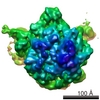
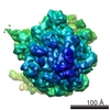
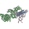
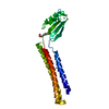
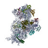
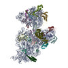
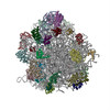
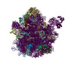
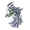
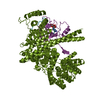
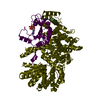
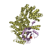
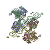
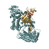
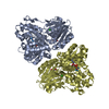
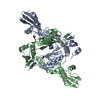
| タイトル | Coordinates of RRF fitted into Cryo-EM map of the 70S post-termination complex | ||||||
 要素 要素 |
| ||||||
 キーワード キーワード | BIOSYNTHETIC/structural protein/RNA / ribosome recycling factor / elongation factor G / 70S / post-termination complex / BIOSYNTHETIC-structural protein-RNA COMPLEX | ||||||
| 機能・相同性 |  機能・相同性情報 機能・相同性情報cytoplasmic translational termination / ribosomal large subunit binding / misfolded RNA binding / Group I intron splicing / RNA folding / positive regulation of RNA splicing / maintenance of translational fidelity / cytosolic small ribosomal subunit / cytoplasmic translation / tRNA binding ...cytoplasmic translational termination / ribosomal large subunit binding / misfolded RNA binding / Group I intron splicing / RNA folding / positive regulation of RNA splicing / maintenance of translational fidelity / cytosolic small ribosomal subunit / cytoplasmic translation / tRNA binding / rRNA binding / structural constituent of ribosome / ribosome / translation / response to antibiotic / cytoplasm / cytosol 類似検索 - 分子機能 | ||||||
| 生物種 |  | ||||||
| 手法 | 電子顕微鏡法 / 単粒子再構成法 / クライオ電子顕微鏡法 / 解像度: 14.1 Å | ||||||
 データ登録者 データ登録者 | Gao, N. / Zavialov, A.V. / Li, W. / Sengupta, J. / Valle, M. / Gursky, R.P. / Ehrenberg, M. / Frank, J. | ||||||
 引用 引用 | ジャーナル: Mol Cell / 年: 2005 タイトル: Mechanism for the disassembly of the posttermination complex inferred from cryo-EM studies. 著者: Ning Gao / Andrey V Zavialov / Wen Li / Jayati Sengupta / Mikel Valle / Richard P Gursky / Måns Ehrenberg / Joachim Frank /  要旨: Ribosome recycling, the disassembly of the posttermination complex after each round of protein synthesis, is an essential step in mRNA translation, but its mechanism has remained obscure. In ...Ribosome recycling, the disassembly of the posttermination complex after each round of protein synthesis, is an essential step in mRNA translation, but its mechanism has remained obscure. In eubacteria, recycling is catalyzed by RRF (ribosome recycling factor) and EF-G (elongation factor G). By using cryo-electron microscopy, we have obtained two density maps, one of the RRF bound posttermination complex and one of the 50S subunit bound with both EF-G and RRF. Comparing the two maps, we found domain I of RRF to be in the same orientation, while domain II in the EF-G-containing 50S subunit is extensively rotated (approximately 60 degrees) compared to its orientation in the 70S complex. Mapping the 50S conformation of RRF onto the 70S posttermination complex suggests that it can disrupt the intersubunit bridges B2a and B3, and thus effect a separation of the two subunits. These observations provide the structural basis for the mechanism by which the posttermination complex is split into subunits by the joint action of RRF and EF-G. #1: ジャーナル: Cell(Cambridge,Mass.) / 年: 2003タイトル: Locking and unlocking of ribosomal motions 著者: Valle, M. / Zavialov, A. / Sengupta, J. / Rawat, U. / Ehrenberg, M. / Frank, J. #2: ジャーナル: Cell(Cambridge,Mass.) / 年: 2003タイトル: Study of structural dynamics of E.coli 70S ribosome using real-space refinement 著者: Gao, H. / Sengupta, J. / Valle, M. / Korostelev, A. / Eswar, N. / Stagg, S.M. / Van Roey, P. / Agrawal, R.K. / Harvey, S.C. / Sali, A. / Chapman, M.S. / Frank, J. #3: ジャーナル: Embo J. / 年: 2000タイトル: Crystal structure of the ribosome recycling factor from E.coli 著者: Kim, K.K. / Min, K. / Suh, S.W. | ||||||
| 履歴 |
| ||||||
| Remark 999 | SEQUENCE This entry contains C alpha and P atoms only. |
- 構造の表示
構造の表示
| ムービー |
ムービービューア |
|---|---|
| 構造ビューア | 分子: MolmilJmol/JSmol |
- ダウンロードとリンク
-ダウンロード
| PDBx/mmCIF形式 | 1zn1.cif.gz | 26.5 KB | 表示 | PDBx/mmCIF形式 |
|---|---|---|---|---|
| PDB形式 | pdb1zn1.ent.gz | 11.3 KB | 表示 | PDB形式 |
| PDBx/mmJSON形式 | 1zn1.json.gz | ツリー表示 | PDBx/mmJSON形式 | |
| その他 |  その他のダウンロード その他のダウンロード |
-検証レポート
| 文書・要旨 | 1zn1_validation.pdf.gz | 839.9 KB | 表示 | wwPDB検証レポート |
|---|---|---|---|---|
| 文書・詳細版 | 1zn1_full_validation.pdf.gz | 839.5 KB | 表示 | |
| XML形式データ | 1zn1_validation.xml.gz | 11.8 KB | 表示 | |
| CIF形式データ | 1zn1_validation.cif.gz | 15.8 KB | 表示 | |
| アーカイブディレクトリ | https://data.pdbj.org/pub/pdb/validation_reports/zn/1zn1ftp://data.pdbj.org/pub/pdb/validation_reports/zn/1zn1 | HTTPS FTP |
-関連構造データ
-リンク
 PDBj
PDBj


- 集合体
集合体
| 登録構造単位 | 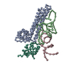
|
|---|---|
| 1 |
|
-要素
| #1: RNA鎖 | 分子量: 18952.248 Da / 分子数: 1 / 断片: fragment of large subunit rRNA helix 69-71 / 由来タイプ: 天然 / 由来: (天然) |
|---|---|
| #2: RNA鎖 | 分子量: 12988.728 Da / 分子数: 1 / 断片: fragment of small subunit rRNA helix 44 / 由来タイプ: 天然 / 由来: (天然) |
| #3: タンパク質 | 分子量: 20671.621 Da / 分子数: 1 / 由来タイプ: 天然 / 由来: (天然) |
| #4: タンパク質 | 分子量: 10828.608 Da / 分子数: 1 / 由来タイプ: 天然 / 由来: (天然) |
-実験情報
-実験
| 実験 | 手法: 電子顕微鏡法 |
|---|---|
| EM実験 | 試料の集合状態: PARTICLE / 3次元再構成法: 単粒子再構成法 |
- 試料調製
試料調製
| 構成要素 | 名称: 70S ribosome / タイプ: RIBOSOME |
|---|---|
| 緩衝液 | 名称: Polymix buffer / pH: 7.5 / 詳細: Polymix buffer |
| 試料 | 濃度: 0.032 mg/ml / 包埋: NO / シャドウイング: NO / 染色: NO / 凍結: YES |
| 試料支持 | 詳細: Quantifoil holey carbon film grids |
| 急速凍結 | 装置: HOMEMADE PLUNGER / 凍結剤: ETHANE / 詳細: Rapid-freezing in liquid ethane |
- 電子顕微鏡撮影
| 実験機器 |  モデル: Tecnai F20 / 画像提供: FEI Company |
|---|---|
| 顕微鏡 | モデル: FEI TECNAI F20 / 日付: 2003年2月1日 |
| 電子銃 | 電子線源:  FIELD EMISSION GUN / 加速電圧: 200 kV / 照射モード: FLOOD BEAM FIELD EMISSION GUN / 加速電圧: 200 kV / 照射モード: FLOOD BEAM |
| 電子レンズ | モード: BRIGHT FIELD / 倍率(公称値): 50000 X / 倍率(補正後): 49700 X / 最大 デフォーカス(公称値): -3.8 nm / 最小 デフォーカス(公称値): -1.4 nm |
| 試料ホルダ | 温度: 93 K / 傾斜角・最大: 0 ° / 傾斜角・最小: 0 ° |
| 撮影 | 電子線照射量: 15 e/Å2 / フィルム・検出器のモデル: KODAK SO-163 FILM |
- 解析
解析
| CTF補正 | 詳細: CTF correction of 3D maps by Wiener filtration | ||||||||||||||||||||||||||||
|---|---|---|---|---|---|---|---|---|---|---|---|---|---|---|---|---|---|---|---|---|---|---|---|---|---|---|---|---|---|
| 対称性 | 点対称性: C1 (非対称) | ||||||||||||||||||||||||||||
| 3次元再構成 | 手法: 3D projection matching; conjugate gradient with regularization 解像度: 14.1 Å / 粒子像の数: 37379 / ピクセルサイズ(実測値): 2.82 Å / 倍率補正: TMV / 詳細: SPIDER package / 対称性のタイプ: POINT | ||||||||||||||||||||||||||||
| 原子モデル構築 | プロトコル: OTHER / 空間: REAL / 詳細: METHOD--Manual fitting in O | ||||||||||||||||||||||||||||
| 原子モデル構築 |
| ||||||||||||||||||||||||||||
| 精密化ステップ | サイクル: LAST
|

