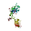
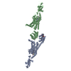
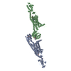
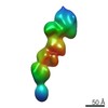
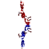
Anosmin1 / Kallmann syndrome protein / Adhesion molecule-like X-linked / Coordinate model: Cα atoms only
Mass: 76545.117 Da / Num. of mol.: 1 Source method: isolated from a genetically manipulated source Source: (gene. exp.) Homo sapiens (human) / Gene: KAL1, ADMLX, KAL, KALIG1 / Plasmid: pMT/BIP/V5-His / Production host: Drosophila melanogaster (fruit fly) / Strain (production host): Schneider 2 (S2) cells / References: UniProt: P23352
-
Experimental details
-
Experiment
Experiment
Method: SOLUTION SCATTERING
-
Data collection
Diffraction
ID
Mean temperature (K)
Crystal-ID
1
288
1
2
1
Diffraction source
Source: SYNCHROTRON / Site: ESRF / Beamline: ID2
Detector
Type
ID
Detector
Date
FRELON CCD CAMERA
1
CCD
May 1, 2003
FRELON CCD CAMERA
2
CCD
Dec 1, 2003
Radiation
Protocol: SINGLE WAVELENGTH / Monochromatic (M) / Laue (L): M / Scattering type: x-ray
Radiation wavelength
Relative weight: 1
Soln scatter
Type: x-ray / Buffer name: 12 MM NA PHOSPHATE, 220 MM NACL / Conc. range: 0.2-0.4 / Data analysis software list: SCTPL7, GNOM / Data reduction software list: MULTICCD / Detector type: FRELON CCD CAMERA / Mean guiner radius: 6.65 nm / Mean guiner radius esd: 0.49 nm / Min mean cross sectional radii gyration: 1.88 nm / Min mean cross sectional radii gyration esd: 0.77 nm / Num. of time frames: 1 / Protein length: 1 / Sample pH: 7.4 / Source beamline: ID02 / Source class: Y / Source type: ESRF BEAMLINE ID02 / Temperature: 288 K
-
Processing
Software
Name
Version
Classification
MULTICCD
datacollection
SCTPL7
datareduction
GNOM
datareduction
Insight II
II98.0
modelbuilding
SCTPL7
modelbuilding
MULTICCD
datareduction
SCTPL7
datascaling
GNOM
datascaling
SCTPL7
phasing
Refinement step
Cycle: LAST
Protein
Nucleic acid
Ligand
Solvent
Total
Num. atoms
680
0
0
0
680
Soln scatter model
Method: CONSTRAINED SCATTERING FITTING OF HOMOLOGY MODELS Conformer selection criteria: THE MODELLED SCATTERING CURVES WERE ASSESSED BY CALCULATION OF THE RG, RSX-1 AND VALUES IN THE SAME Q RANGES USED IN THE EXPERIMENTAL GUINIER FITS. MODELS WERE THEN ...Conformer selection criteria: THE MODELLED SCATTERING CURVES WERE ASSESSED BY CALCULATION OF THE RG, RSX-1 AND VALUES IN THE SAME Q RANGES USED IN THE EXPERIMENTAL GUINIER FITS. MODELS WERE THEN RANKED USING A GOODNESS-OF-FIT R-FACTOR DEFINED BY ANALOGY WITH PROTEIN CRYSTALLOGRAPHY Details: HOMOLOGY MODELS WERE BUILT FOR THE CYS-BOX, WAP AND FOUR FNIII DOMAINS. THE POSITIONS OF THE DOMAINS WERE DETERMINED BY AN APPROACH THAT COMBINED RANDOMISED LINKER PEPTIDE STRUCTURES ...Details: HOMOLOGY MODELS WERE BUILT FOR THE CYS-BOX, WAP AND FOUR FNIII DOMAINS. THE POSITIONS OF THE DOMAINS WERE DETERMINED BY AN APPROACH THAT COMBINED RANDOMISED LINKER PEPTIDE STRUCTURES PRODUCED BY MOLECULAR DYNAMICS SIMULATIONS WITH CURVE-FITTING TO EXPERIMENTAL X-RAY SOLUTION SCATTERING DATA. A SINGLE ARRANGEMENT OF THE SIX DOMAINS IS PRESENTED, WHICH IS REPRESENTATIVE OF A FAMILY OF STRUCTURES THAT FIT THE SCATTERING DATA. MORE DETAILS ON THE MODELLING STRATEGY ARE CONTAINED IN THE PRIMARY REFERENCE. THE ALGORITHM RESULTED IN SEVERAL CLOSE APPROACHES BETWEEN ATOMS. THIS ENTRY CONTAINS FOUR SUCH PAIRS ALA 292 - PRO 318 PRO 291 - GLU 319 PRO 27 - PHE 122 TYR 225 - PHE 355 Num. of conformers calculated: 10000 / Num. of conformers submitted: 1 / Representative conformer: 1 / Software author list: ACCELRYS Software list: INSIGHT II, HOMOLOGY, DISCOVERY, BIOPOLYMER, DELPHI, O, SCTPL7, GNOM
+
About Yorodumi
-
News
-
Feb 9, 2022. New format data for meta-information of EMDB entries
New format data for meta-information of EMDB entries
Version 3 of the EMDB header file is now the official format.
The previous official version 1.9 will be removed from the archive.
In the structure databanks used in Yorodumi, some data are registered as the other names, "COVID-19 virus" and "2019-nCoV". Here are the details of the virus and the list of structure data.
Jan 31, 2019. EMDB accession codes are about to change! (news from PDBe EMDB page)
EMDB accession codes are about to change! (news from PDBe EMDB page)
The allocation of 4 digits for EMDB accession codes will soon come to an end. Whilst these codes will remain in use, new EMDB accession codes will include an additional digit and will expand incrementally as the available range of codes is exhausted. The current 4-digit format prefixed with “EMD-” (i.e. EMD-XXXX) will advance to a 5-digit format (i.e. EMD-XXXXX), and so on. It is currently estimated that the 4-digit codes will be depleted around Spring 2019, at which point the 5-digit format will come into force.
The EM Navigator/Yorodumi systems omit the EMD- prefix.
Related info.:Q: What is EMD? / ID/Accession-code notation in Yorodumi/EM Navigator
Yorodumi is a browser for structure data from EMDB, PDB, SASBDB, etc.
This page is also the successor to EM Navigator detail page, and also detail information page/front-end page for Omokage search.
The word "yorodu" (or yorozu) is an old Japanese word meaning "ten thousand". "mi" (miru) is to see.
Related info.:EMDB / PDB / SASBDB / Comparison of 3 databanks / Yorodumi Search / Aug 31, 2016. New EM Navigator & Yorodumi / Yorodumi Papers / Jmol/JSmol / Function and homology information / Changes in new EM Navigator and Yorodumi
 Movie
Movie Controller


 Open data
 Basic information
 Components
 Keywords
 Function and homology information
 Homo sapiens (human)
 SOLUTION SCATTERING /
  Authors
 Citation
 Structure visualization
 Downloads & links
 Other downloads
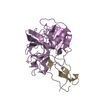


 PDBj
PDBj


 Assembly
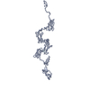

 / Beamline: ID2
 Processing