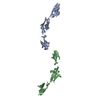
 Movie
Movie Controller


+ Open data

- Basic information
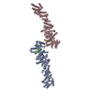
| Entry | Database: PDB / ID: 4fom | |||||||||
|---|---|---|---|---|---|---|---|---|---|---|
| Title | Crystal structure of human nectin-3 full ectodomain (D1-D3) | |||||||||
 Components Components | Poliovirus receptor-related protein 3 | |||||||||
 Keywords Keywords | CELL ADHESION / Immunoglobulin-like domain / Ig domain | |||||||||
| Function / homology |  Function and homology information Function and homology informationretina morphogenesis in camera-type eye / Nectin/Necl trans heterodimerization / establishment of protein localization to plasma membrane / protein localization to cell junction / lens morphogenesis in camera-type eye / cell adhesion mediator activity / cochlea morphogenesis / cell-cell contact zone / fertilization / Adherens junctions interactions ...retina morphogenesis in camera-type eye / Nectin/Necl trans heterodimerization / establishment of protein localization to plasma membrane / protein localization to cell junction / lens morphogenesis in camera-type eye / cell adhesion mediator activity / cochlea morphogenesis / cell-cell contact zone / fertilization / Adherens junctions interactions / heterophilic cell-cell adhesion / apical junction complex / homophilic cell-cell adhesion / regulation of postsynapse assembly / cell adhesion molecule binding / hippocampal mossy fiber to CA3 synapse / adherens junction / postsynaptic density membrane / cell-cell junction / axon / symbiont entry into host cell / dendrite / protein homodimerization activity / identical protein binding / plasma membrane Similarity search - Function | |||||||||
| Biological species |  Homo sapiens (human) Homo sapiens (human) | |||||||||
| Method |  X-RAY DIFFRACTION / SYNCHROTRON / MOLECULAR REPLACEMENT / Resolution: 3.93 Å X-RAY DIFFRACTION / SYNCHROTRON / MOLECULAR REPLACEMENT / Resolution: 3.93 Å | |||||||||
 Authors Authors | Harrison, O.J. / Jin, X. / Brasch, J. / Shapiro, L. | |||||||||
 Citation Citation | Journal: Nat.Struct.Mol.Biol. / Year: 2012 Title: Nectin ectodomain structures reveal a canonical adhesive interface. Authors: Harrison, O.J. / Vendome, J. / Brasch, J. / Jin, X. / Hong, S. / Katsamba, P.S. / Ahlsen, G. / Troyanovsky, R.B. / Troyanovsky, S.M. / Honig, B. / Shapiro, L. | |||||||||
| History |
|
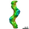
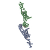
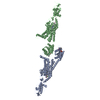
- Structure visualization
| Structure viewer | Molecule: MolmilJmol/JSmol |
|---|
- Downloads & links
-Download
| PDBx/mmCIF format | 4fom.cif.gz | 144.2 KB | Display | PDBx/mmCIF format |
|---|---|---|---|---|
| PDB format | pdb4fom.ent.gz | 115.3 KB | Display | PDB format |
| PDBx/mmJSON format | 4fom.json.gz | Tree view | PDBx/mmJSON format | |
| Others |  Other downloads Other downloads |
-Validation report
| Arichive directory | https://data.pdbj.org/pub/pdb/validation_reports/fo/4fomftp://data.pdbj.org/pub/pdb/validation_reports/fo/4fom | HTTPS FTP |
|---|
-Related structure data
| Related structure data |  4fmfC  4fmkC  4fn0C  4fqpC 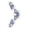 4frwC  4fs0C  3alpS C: citing same article ( S: Starting model for refinement |
|---|---|
| Similar structure data |

-Links
 PDBj
PDBj


- Assembly
| Deposited unit | 
| ||||||||
|---|---|---|---|---|---|---|---|---|---|
| 1 | 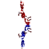
| ||||||||
| Unit cell |
|
-Components
| #1: Protein | Mass: 34039.387 Da / Num. of mol.: 1 / Fragment: ectodomain (D1-D3, UNP residues 58-359) Source method: isolated from a genetically manipulated source Source: (gene. exp.) Homo sapiens (human) / Gene: PVRL3, PRR3 / Plasmid: pCEP4 / Cell line (production host): HEK 293F / Production host: Homo sapiens (human) / References: UniProt: Q9NQS3 | ||||||||
|---|---|---|---|---|---|---|---|---|---|
| #2: Polysaccharide | Source method: isolated from a genetically manipulated source #3: Polysaccharide | beta-D-mannopyranose-(1-4)-2-acetamido-2-deoxy-beta-D-glucopyranose-(1-4)-2-acetamido-2-deoxy-beta- ...beta-D-mannopyranose-(1-4)-2-acetamido-2-deoxy-beta-D-glucopyranose-(1-4)-2-acetamido-2-deoxy-beta-D-glucopyranose | Source method: isolated from a genetically manipulated source #4: Polysaccharide | alpha-D-mannopyranose-(1-3)-[alpha-D-mannopyranose-(1-6)]beta-D-mannopyranose-(1-4)-2-acetamido-2- ...alpha-D-mannopyranose-(1-3)-[alpha-D-mannopyranose-(1-6)]beta-D-mannopyranose-(1-4)-2-acetamido-2-deoxy-beta-D-glucopyranose-(1-4)-[alpha-L-fucopyranose-(1-6)]2-acetamido-2-deoxy-beta-D-glucopyranose | Source method: isolated from a genetically manipulated source #5: Sugar | ChemComp-NAG / |   Type: D-saccharide, beta linking / Mass: 221.208 Da / Num. of mol.: 1 Type: D-saccharide, beta linking / Mass: 221.208 Da / Num. of mol.: 1Source method: isolated from a genetically manipulated source Formula: C8H15NO6 Has protein modification | Y | |
-Experimental details
-Experiment
| Experiment | Method: X-RAY DIFFRACTION / Number of used crystals: 1 |
|---|
- Sample preparation
| Crystal | Density Matthews: 9.13 Å3/Da / Density % sol: 86.52 % |
|---|---|
| Crystal grow | Temperature: 293.15 K / Method: vapor diffusion, hanging drop / pH: 4.6 Details: 4.6 M ammonium acetate, 0.1 M sodium acetate, VAPOR DIFFUSION, HANGING DROP, temperature 293.15K |
-Data collection
| Diffraction | Mean temperature: 100 K | ||||||||||||||||||||||||||||||||||||||||||
|---|---|---|---|---|---|---|---|---|---|---|---|---|---|---|---|---|---|---|---|---|---|---|---|---|---|---|---|---|---|---|---|---|---|---|---|---|---|---|---|---|---|---|---|
| Diffraction source | Source: SYNCHROTRON / Site: NSLS  / Beamline: X4C / Wavelength: 0.9792 Å / Beamline: X4C / Wavelength: 0.9792 Å | ||||||||||||||||||||||||||||||||||||||||||
| Detector | Type: MAR CCD 165 mm / Detector: CCD / Date: Jul 1, 2011 | ||||||||||||||||||||||||||||||||||||||||||
| Radiation | Monochromator: Bent single Si(111) crystal (horizontal focusing and deflection) Protocol: SINGLE WAVELENGTH / Monochromatic (M) / Laue (L): M / Scattering type: x-ray | ||||||||||||||||||||||||||||||||||||||||||
| Radiation wavelength | Wavelength: 0.9792 Å / Relative weight: 1 | ||||||||||||||||||||||||||||||||||||||||||
| Reflection | Resolution: 3.93→40 Å / Num. all: 11776 / Num. obs: 11776 / % possible obs: 98.3 % / Observed criterion σ(I): -3 / Redundancy: 5.9 % / Rsym value: 0.07 / Net I/σ(I): 34 | ||||||||||||||||||||||||||||||||||||||||||
| Reflection shell |
|
- Processing
| Software |
| ||||||||||||||||||||||||||||||||||||||||||||||||||||||||||||||||||||||||||||||||||||||||||||||||||||
|---|---|---|---|---|---|---|---|---|---|---|---|---|---|---|---|---|---|---|---|---|---|---|---|---|---|---|---|---|---|---|---|---|---|---|---|---|---|---|---|---|---|---|---|---|---|---|---|---|---|---|---|---|---|---|---|---|---|---|---|---|---|---|---|---|---|---|---|---|---|---|---|---|---|---|---|---|---|---|---|---|---|---|---|---|---|---|---|---|---|---|---|---|---|---|---|---|---|---|---|---|---|
| Refinement | Method to determine structure: MOLECULAR REPLACEMENT Starting model: PDB ENTRY 3ALP Resolution: 3.93→20 Å / Cor.coef. Fo:Fc: 0.932 / Cor.coef. Fo:Fc free: 0.907 / SU B: 62.326 / SU ML: 0.467 / Cross valid method: THROUGHOUT / ESU R: 1.721 / ESU R Free: 0.538 / Stereochemistry target values: MAXIMUM LIKELIHOOD / Details: HYDROGENS HAVE BEEN ADDED IN THE RIDING POSITIONS
| ||||||||||||||||||||||||||||||||||||||||||||||||||||||||||||||||||||||||||||||||||||||||||||||||||||
| Solvent computation | Ion probe radii: 0.8 Å / Shrinkage radii: 0.8 Å / VDW probe radii: 1.2 Å / Solvent model: MASK | ||||||||||||||||||||||||||||||||||||||||||||||||||||||||||||||||||||||||||||||||||||||||||||||||||||
| Displacement parameters | Biso mean: 170.513 Å2
| ||||||||||||||||||||||||||||||||||||||||||||||||||||||||||||||||||||||||||||||||||||||||||||||||||||
| Refinement step | Cycle: LAST / Resolution: 3.93→20 Å
| ||||||||||||||||||||||||||||||||||||||||||||||||||||||||||||||||||||||||||||||||||||||||||||||||||||
| Refine LS restraints |
| ||||||||||||||||||||||||||||||||||||||||||||||||||||||||||||||||||||||||||||||||||||||||||||||||||||
| LS refinement shell | Resolution: 3.93→4.029 Å / Total num. of bins used: 20
| ||||||||||||||||||||||||||||||||||||||||||||||||||||||||||||||||||||||||||||||||||||||||||||||||||||
| Refinement TLS params. | Method: refined / Refine-ID: X-RAY DIFFRACTION
| ||||||||||||||||||||||||||||||||||||||||||||||||||||||||||||||||||||||||||||||||||||||||||||||||||||
| Refinement TLS group |
|
