 Movie
Movie Controller

[English] 日本語
 Yorodumi
- PDB-4fs0: Crystal structure of mutant F136D of mouse nectin-2 extracellular... -
+ Open data

- Basic information
| Entry | Database: PDB / ID: 4fs0 | |||||||||
|---|---|---|---|---|---|---|---|---|---|---|
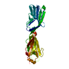
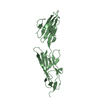
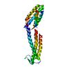
| Title | Crystal structure of mutant F136D of mouse nectin-2 extracellular fragment D1-D2 | |||||||||
 Components Components | Poliovirus receptor-related protein 2 | |||||||||
 Keywords Keywords | CELL ADHESION / Immunoglobulin-like domain / Ig domain / viral entry receptor | |||||||||
| Function / homology |  Function and homology information Function and homology informationcellular anatomical entity morphogenesis / Nectin/Necl trans heterodimerization / coreceptor-mediated virion attachment to host cell / establishment of mitochondrion localization / sperm mitochondrion organization / positive regulation of immunoglobulin mediated immune response / susceptibility to T cell mediated cytotoxicity / susceptibility to natural killer cell mediated cytotoxicity / spermatid nucleus differentiation / regulation of viral entry into host cell ...cellular anatomical entity morphogenesis / Nectin/Necl trans heterodimerization / coreceptor-mediated virion attachment to host cell / establishment of mitochondrion localization / sperm mitochondrion organization / positive regulation of immunoglobulin mediated immune response / susceptibility to T cell mediated cytotoxicity / susceptibility to natural killer cell mediated cytotoxicity / spermatid nucleus differentiation / regulation of viral entry into host cell / positive regulation of mast cell activation / positive regulation of natural killer cell mediated cytotoxicity directed against tumor cell target / Adherens junctions interactions / acrosome assembly / adhesion of symbiont to host / zonula adherens / cilium organization / cell-cell contact zone / positive regulation of T cell receptor signaling pathway / positive regulation of natural killer cell mediated cytotoxicity / negative regulation of natural killer cell mediated cytotoxicity / fertilization / Immunoregulatory interactions between a Lymphoid and a non-Lymphoid cell / natural killer cell mediated cytotoxicity / apical junction complex / homophilic cell-cell adhesion / spermatid development / cytoskeleton organization / cell adhesion molecule binding / cell-cell junction / receptor ligand activity / fusion of virus membrane with host plasma membrane / cell surface / protein homodimerization activity / membrane / identical protein binding / plasma membrane Similarity search - Function | |||||||||
| Biological species |  | |||||||||
| Method |  X-RAY DIFFRACTION / SYNCHROTRON / MOLECULAR REPLACEMENT / Resolution: 3.25 Å X-RAY DIFFRACTION / SYNCHROTRON / MOLECULAR REPLACEMENT / Resolution: 3.25 Å | |||||||||
 Authors Authors | Harrison, O.J. / Brasch, J. / Shapiro, L. | |||||||||
 Citation Citation | Journal: Nat.Struct.Mol.Biol. / Year: 2012 Title: Nectin ectodomain structures reveal a canonical adhesive interface. Authors: Harrison, O.J. / Vendome, J. / Brasch, J. / Jin, X. / Hong, S. / Katsamba, P.S. / Ahlsen, G. / Troyanovsky, R.B. / Troyanovsky, S.M. / Honig, B. / Shapiro, L. | |||||||||
| History |
|
- Structure visualization
| Structure viewer | Molecule: MolmilJmol/JSmol |
|---|
- Downloads & links
-Download
| PDBx/mmCIF format | 4fs0.cif.gz | 96.4 KB | Display | PDBx/mmCIF format |
|---|---|---|---|---|
| PDB format | pdb4fs0.ent.gz | 73.4 KB | Display | PDB format |
| PDBx/mmJSON format | 4fs0.json.gz | Tree view | PDBx/mmJSON format | |
| Others |  Other downloads Other downloads |
-Validation report
| Arichive directory | https://data.pdbj.org/pub/pdb/validation_reports/fs/4fs0ftp://data.pdbj.org/pub/pdb/validation_reports/fs/4fs0 | HTTPS FTP |
|---|
-Related structure data
| Related structure data |  4fmfC  4fmkSC  4fn0C  4fomC  4fqpC  4frwC C: citing same article ( S: Starting model for refinement |
|---|---|
| Similar structure data |

-Links
 PDBj
PDBj


- Assembly
| Deposited unit | 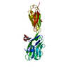
| ||||||||
|---|---|---|---|---|---|---|---|---|---|
| 1 |
| ||||||||
| Unit cell |
| ||||||||
| Components on special symmetry positions |
|
-Components
| #1: Protein | Mass: 24887.990 Da / Num. of mol.: 1 / Fragment: extracellular domain (D1-D2, UNP residues 32-250) / Mutation: F136D Source method: isolated from a genetically manipulated source Source: (gene. exp.)  Homo sapiens (human) / References: UniProt: P32507 Homo sapiens (human) / References: UniProt: P32507 |
|---|---|
| #2: Polysaccharide | 2-acetamido-2-deoxy-beta-D-glucopyranose-(1-4)-[alpha-L-fucopyranose-(1-6)]2-acetamido-2-deoxy-beta- ...2-acetamido-2-deoxy-beta-D-glucopyranose-(1-4)-[alpha-L-fucopyranose-(1-6)]2-acetamido-2-deoxy-beta-D-glucopyranose Source method: isolated from a genetically manipulated source |
| #3: Chemical | ChemComp-SO4 /   Mass: 96.063 Da / Num. of mol.: 1 / Source method: obtained synthetically / Formula: SO4 Mass: 96.063 Da / Num. of mol.: 1 / Source method: obtained synthetically / Formula: SO4 |
| #4: Water | ChemComp-HOH /  Mass: 18.015 Da / Num. of mol.: 12 / Source method: isolated from a natural source / Formula: H2O Mass: 18.015 Da / Num. of mol.: 12 / Source method: isolated from a natural source / Formula: H2O |
| Has protein modification | Y |
-Experimental details
-Experiment
| Experiment | Method: X-RAY DIFFRACTION / Number of used crystals: 1 |
|---|
- Sample preparation
| Crystal | Density Matthews: 2.18 Å3/Da / Density % sol: 43.63 % |
|---|---|
| Crystal grow | Temperature: 293.15 K / Method: vapor diffusion, hanging drop / pH: 5.5 Details: 1 M lithium sulfate, 0.6 M ammonium sulfate, 0.1 M tri-sodium citrate, pH 5.5, cryoprotectant: 30% w/v glycerol, VAPOR DIFFUSION, HANGING DROP, temperature 293.15K |
-Data collection
| Diffraction | Mean temperature: 100 K | ||||||||||||||||||||||||||||||||||||||||||||||||||||||||||||||||||
|---|---|---|---|---|---|---|---|---|---|---|---|---|---|---|---|---|---|---|---|---|---|---|---|---|---|---|---|---|---|---|---|---|---|---|---|---|---|---|---|---|---|---|---|---|---|---|---|---|---|---|---|---|---|---|---|---|---|---|---|---|---|---|---|---|---|---|---|
| Diffraction source | Source: SYNCHROTRON / Site: NSLS  / Beamline: X4C / Wavelength: 0.9792 Å / Beamline: X4C / Wavelength: 0.9792 Å | ||||||||||||||||||||||||||||||||||||||||||||||||||||||||||||||||||
| Detector | Type: MAR CCD 165 mm / Detector: CCD / Date: Mar 3, 2012 | ||||||||||||||||||||||||||||||||||||||||||||||||||||||||||||||||||
| Radiation | Monochromator: Bent single Si(111) crystal (horizontal focusing and deflection) Protocol: SINGLE WAVELENGTH / Monochromatic (M) / Laue (L): M / Scattering type: x-ray | ||||||||||||||||||||||||||||||||||||||||||||||||||||||||||||||||||
| Radiation wavelength | Wavelength: 0.9792 Å / Relative weight: 1 | ||||||||||||||||||||||||||||||||||||||||||||||||||||||||||||||||||
| Reflection | Resolution: 3.25→30 Å / Num. all: 19387 / Num. obs: 19387 / % possible obs: 99.8 % / Observed criterion σ(I): -3 / Redundancy: 6.3 % / Rsym value: 0.11 / Net I/σ(I): 13.7 | ||||||||||||||||||||||||||||||||||||||||||||||||||||||||||||||||||
| Reflection shell |
|
- Processing
| Software |
| ||||||||||||||||||||||||||||||||||||||||||||||||||||||||||||||||||||||||||||||||||||||||||||||||||||||||||||||||||||||||||||||||||||||||||||||||||||||||||||||||||||||||||
|---|---|---|---|---|---|---|---|---|---|---|---|---|---|---|---|---|---|---|---|---|---|---|---|---|---|---|---|---|---|---|---|---|---|---|---|---|---|---|---|---|---|---|---|---|---|---|---|---|---|---|---|---|---|---|---|---|---|---|---|---|---|---|---|---|---|---|---|---|---|---|---|---|---|---|---|---|---|---|---|---|---|---|---|---|---|---|---|---|---|---|---|---|---|---|---|---|---|---|---|---|---|---|---|---|---|---|---|---|---|---|---|---|---|---|---|---|---|---|---|---|---|---|---|---|---|---|---|---|---|---|---|---|---|---|---|---|---|---|---|---|---|---|---|---|---|---|---|---|---|---|---|---|---|---|---|---|---|---|---|---|---|---|---|---|---|---|---|---|---|---|---|
| Refinement | Method to determine structure: MOLECULAR REPLACEMENT Starting model: PDB ENTRY 4FMK Resolution: 3.25→20 Å / Cor.coef. Fo:Fc: 0.896 / Cor.coef. Fo:Fc free: 0.789 / SU B: 77.143 / SU ML: 0.641 / Cross valid method: THROUGHOUT / ESU R Free: 0.794 / Stereochemistry target values: MAXIMUM LIKELIHOOD / Details: HYDROGENS HAVE BEEN ADDED IN THE RIDING POSITIONS
| ||||||||||||||||||||||||||||||||||||||||||||||||||||||||||||||||||||||||||||||||||||||||||||||||||||||||||||||||||||||||||||||||||||||||||||||||||||||||||||||||||||||||||
| Solvent computation | Ion probe radii: 0.8 Å / Shrinkage radii: 0.8 Å / VDW probe radii: 1.2 Å / Solvent model: MASK | ||||||||||||||||||||||||||||||||||||||||||||||||||||||||||||||||||||||||||||||||||||||||||||||||||||||||||||||||||||||||||||||||||||||||||||||||||||||||||||||||||||||||||
| Displacement parameters | Biso mean: 52.836 Å2
| ||||||||||||||||||||||||||||||||||||||||||||||||||||||||||||||||||||||||||||||||||||||||||||||||||||||||||||||||||||||||||||||||||||||||||||||||||||||||||||||||||||||||||
| Refinement step | Cycle: LAST / Resolution: 3.25→20 Å
| ||||||||||||||||||||||||||||||||||||||||||||||||||||||||||||||||||||||||||||||||||||||||||||||||||||||||||||||||||||||||||||||||||||||||||||||||||||||||||||||||||||||||||
| Refine LS restraints |
| ||||||||||||||||||||||||||||||||||||||||||||||||||||||||||||||||||||||||||||||||||||||||||||||||||||||||||||||||||||||||||||||||||||||||||||||||||||||||||||||||||||||||||
| LS refinement shell | Resolution: 3.25→3.335 Å / Total num. of bins used: 20
| ||||||||||||||||||||||||||||||||||||||||||||||||||||||||||||||||||||||||||||||||||||||||||||||||||||||||||||||||||||||||||||||||||||||||||||||||||||||||||||||||||||||||||
| Refinement TLS params. | Method: refined / Refine-ID: X-RAY DIFFRACTION
| ||||||||||||||||||||||||||||||||||||||||||||||||||||||||||||||||||||||||||||||||||||||||||||||||||||||||||||||||||||||||||||||||||||||||||||||||||||||||||||||||||||||||||
| Refinement TLS group |
|
