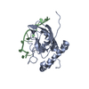
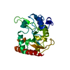
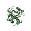
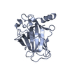
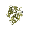
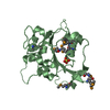
Entry Database : PDB / ID : 1za4Title Crystal Structure of the Thrombospondin-1 N-terminal Domain in Complex with Arixtra Thrombospondin 1 Keywords / / / / Function / homology Function Domain/homology Component
/ / / / / / / / / / / / / / / / / / / / / / / / / / / / / / / / / / / / / / / / / / / / / / / / / / / / / / / / / / / / / / / / / / / / / / / / / / / / / / / / / / / / / / / / / / / / / / / / / / / / / / / / / / / / / / / / / / / / / / / / / / / / / / / / / / / / / / / / / / / / / / / / / / / Biological species Homo sapiens (human)Method / / / Resolution : 1.9 Å Authors Tan, K. / Wang, J.H. / Lawler, J. Journal : Structure / Year : 2006Title : The Structures of the Thrombospondin-1 N-terminal Domain and its Complex with a Synthetic Pentameric HeparinAuthors : Tan, K. / Duquette, M. / Liu, J.H. / Zhang, R. / Joachimiak, A. / Wang, J.H. / Lawler, J. History Deposition Apr 5, 2005 Deposition site / Processing site Revision 1.0 Jan 24, 2006 Provider / Type Revision 1.1 Apr 30, 2008 Group Revision 1.2 Jul 13, 2011 Group Revision 2.0 Jul 29, 2020 Group Atomic model / Data collection ... Atomic model / Data collection / Database references / Derived calculations / Structure summary Category atom_site / chem_comp ... atom_site / chem_comp / entity / pdbx_chem_comp_identifier / pdbx_entity_nonpoly / struct_conn / struct_ref_seq_dif / struct_site / struct_site_gen Item _atom_site.auth_atom_id / _atom_site.label_atom_id ... _atom_site.auth_atom_id / _atom_site.label_atom_id / _chem_comp.mon_nstd_flag / _chem_comp.name / _chem_comp.type / _entity.pdbx_description / _pdbx_entity_nonpoly.name / _struct_conn.pdbx_leaving_atom_flag / _struct_ref_seq_dif.details Description / Provider / Type Revision 2.1 Nov 6, 2024 Group / Database references / Structure summaryCategory chem_comp / chem_comp_atom ... chem_comp / chem_comp_atom / chem_comp_bond / database_2 / pdbx_entry_details / pdbx_modification_feature Item / _database_2.pdbx_DOI / _database_2.pdbx_database_accession
Show all Show less Remark 600 HETEROGEN ARIXTRA (FONDAPARINUX SODIUM) IS A SYNTHETIC MODIFIED PENTAMERIC HEPARIN. THE COMPLETE ... HETEROGEN ARIXTRA (FONDAPARINUX SODIUM) IS A SYNTHETIC MODIFIED PENTAMERIC HEPARIN. THE COMPLETE ARIXTRA MOLECULE WAS NOT OBSERVED. SGN, N,O6-DISULFO-GLUCOSAMINE IS PART OF ARIXTRA MOLECULE (DEFINED AS FIFTH UNIT) AND SO4, SULFATE IONS ARE ALSO PART OF ARIXTRA MOLECULE (FRAGMENTS FROM THE REST OF THE UNITS).
 Movie
Movie Controller

 Yorodumi
 Open data
 Basic information
 Components
 Keywords
 Function and homology information
 Homo sapiens (human)
 X-RAY DIFFRACTION /
  Authors
 Citation
 Structure visualization
 Downloads & links
 Other downloads
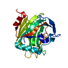
 PDBj
PDBj


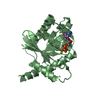
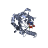
 Assembly
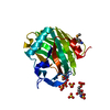

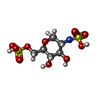
 Type: D-saccharide, alpha linking / Mass: 339.298 Da / Num. of mol.: 1
 Mass: 96.063 Da / Num. of mol.: 5 / Source method: obtained synthetically / Formula: SO4
 Mass: 18.015 Da / Num. of mol.: 78 / Source method: isolated from a natural source / Formula: H2O
 Sample preparation
 Processing