CST complex / ribonucleoprotein complex localization / translation elongation factor binding / telomerase inhibitor activity / regulation of telomere maintenance via telomerase / single-stranded telomeric DNA binding / G-rich strand telomeric DNA binding / nuclear telomere cap complex / telomere capping / telomere maintenance via telomerase ...CST complex / ribonucleoprotein complex localization / translation elongation factor binding / telomerase inhibitor activity / regulation of telomere maintenance via telomerase / single-stranded telomeric DNA binding / G-rich strand telomeric DNA binding / nuclear telomere cap complex / telomere capping / telomere maintenance via telomerase / telomere maintenance / chromosome, telomeric region / cell division / identical protein binding Similarity search - Function
Cell division control protein 13, OB2 domain / Cell division control protein 13, OB2 domain / Cell division control protein 13, N-terminal / Cdc13, OB4 dimerization domain / Cell division control protein 13 N-terminus / Cdc13 OB4 dimerization domain / Protection of telomeres protein 1 / Telomeric single stranded DNA binding POT1/Cdc13 / Telomeric single stranded DNA binding POT1/CDC13 / Telomeric single stranded DNA binding POT1/CDC13 ...Cell division control protein 13, OB2 domain / Cell division control protein 13, OB2 domain / Cell division control protein 13, N-terminal / Cdc13, OB4 dimerization domain / Cell division control protein 13 N-terminus / Cdc13 OB4 dimerization domain / Protection of telomeres protein 1 / Telomeric single stranded DNA binding POT1/Cdc13 / Telomeric single stranded DNA binding POT1/CDC13 / Telomeric single stranded DNA binding POT1/CDC13 / Nucleic acid-binding proteins / OB fold (Dihydrolipoamide Acetyltransferase, E2P) / Nucleic acid-binding, OB-fold / Beta Barrel / Mainly Beta Similarity search - Domain/homology
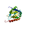
structures with acceptable covalent geometry, structures with the lowest energy
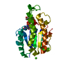
Representative
Model #1
closest to the average
-
Components
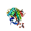
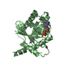
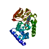
#1: DNA chain
5'-D(*GP*TP*GP*TP*GP*GP*GP*TP*GP*TP*G)-3'
Mass: 3476.254 Da / Num. of mol.: 1 / Source method: obtained synthetically
#2: Protein
Celldivisioncontrolprotein13
Mass: 23575.127 Da / Num. of mol.: 1 / Fragment: DNA-binding domain Source method: isolated from a genetically manipulated source Source: (gene. exp.) Saccharomyces cerevisiae (brewer's yeast) Gene: CDC13, YDL220C / Production host: Escherichia coli (E. coli) / References: UniProt: P32797
-
Experimental details
-
Experiment
Experiment
Method: SOLUTION NMR
NMR experiment
Conditions-ID
Experiment-ID
Solution-ID
Type
1
1
1
2D NOESY
1
2
2
3D 15N-separated NOESY
1
3
3
3D 13C-separated NOESY
1
4
4
4D 13C/15N-separated NOESY
1
5
5
4D 13C-separated NOESY
1
6
6
Isotope filtered NOESY
1
7
7
Select-filtered NOESY
NMR details
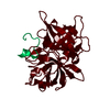
Text: Models superimposed using Douglas Theobald's THESEUS program for multiple superpositions, variance-weighted simultaneous superpositioning. The final weighted RMSD from the mean is 0.64152A over ...Text: Models superimposed using Douglas Theobald's THESEUS program for multiple superpositions, variance-weighted simultaneous superpositioning. The final weighted RMSD from the mean is 0.64152A over all alpha-carbon atoms
-
Sample preparation
Details
Contents: 0.8-1.5mM unlabeled, 15N-labeled or 13C,15N-labeled protein; 0.9-1.7mM ssDNA; 50mM imidazole-d4, pH or pD* 7.0; 150mM NaCl; 100mM Na2SO4; 0.02% NaN3; 2mM DTT-d10 in 10% D2O/90% H2O or 100% D2O Solvent system: 10% D2O/90% H2O or 100% D2O
Conformer selection criteria: structures with acceptable covalent geometry, structures with the lowest energy Conformers calculated total number: 100 / Conformers submitted total number: 10
+
About Yorodumi
-
News
-
Feb 9, 2022. New format data for meta-information of EMDB entries
New format data for meta-information of EMDB entries
Version 3 of the EMDB header file is now the official format.
The previous official version 1.9 will be removed from the archive.
In the structure databanks used in Yorodumi, some data are registered as the other names, "COVID-19 virus" and "2019-nCoV". Here are the details of the virus and the list of structure data.
Jan 31, 2019. EMDB accession codes are about to change! (news from PDBe EMDB page)
EMDB accession codes are about to change! (news from PDBe EMDB page)
The allocation of 4 digits for EMDB accession codes will soon come to an end. Whilst these codes will remain in use, new EMDB accession codes will include an additional digit and will expand incrementally as the available range of codes is exhausted. The current 4-digit format prefixed with “EMD-” (i.e. EMD-XXXX) will advance to a 5-digit format (i.e. EMD-XXXXX), and so on. It is currently estimated that the 4-digit codes will be depleted around Spring 2019, at which point the 5-digit format will come into force.
The EM Navigator/Yorodumi systems omit the EMD- prefix.
Related info.:Q: What is EMD? / ID/Accession-code notation in Yorodumi/EM Navigator
Yorodumi is a browser for structure data from EMDB, PDB, SASBDB, etc.
This page is also the successor to EM Navigator detail page, and also detail information page/front-end page for Omokage search.
The word "yorodu" (or yorozu) is an old Japanese word meaning "ten thousand". "mi" (miru) is to see.
Related info.:EMDB / PDB / SASBDB / Comparison of 3 databanks / Yorodumi Search / Aug 31, 2016. New EM Navigator & Yorodumi / Yorodumi Papers / Jmol/JSmol / Function and homology information / Changes in new EM Navigator and Yorodumi
 Movie
Movie Controller

 Yorodumi
 Open data
 Basic information
 Components
 Keywords
 Function and homology information
 Authors
 Citation
 Structure visualization
 Downloads & links
 Other downloads


 PDBj
PDBj


 Assembly
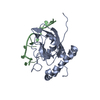

 Sample preparation
 Processing
 X-PLOR