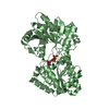
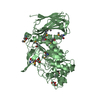
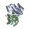
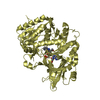
- PDB-5aq6: Structure of E. coli ZinT at 1.79 Angstrom -
+
Open data
ID or keywords:
Loading...
-
Basic information
Entry
Database: PDB / ID: 5aq6
Title
Structure of E. coli ZinT at 1.79 Angstrom
Components
METAL-BINDING PROTEIN ZINT
Keywords
METAL BINDING PROTEIN / ZINC TRANSPORT / NATURAL HIS-TAG / METAL RESISTANCE
Function / homology
Function and homology information
cellular response to zinc ion starvation / intracellular zinc ion homeostasis / cadmium ion binding / cellular response to cadmium ion / cellular response to hydrogen peroxide / outer membrane-bounded periplasmic space / zinc ion binding / metal ion binding / cytosol Similarity search - Function
Protocol: SINGLE WAVELENGTH / Monochromatic (M) / Laue (L): M / Scattering type: x-ray
Radiation wavelength
Wavelength: 2.1 Å / Relative weight: 1
Reflection
Resolution: 1.79→45.8 Å / Num. obs: 28379 / % possible obs: 99.6 % / Observed criterion σ(I): 1.7 / Redundancy: 6.7 % / Biso Wilson estimate: 24.73 Å2 / Rmerge(I) obs: 0.07 / Net I/σ(I): 15.6
Reflection shell
Resolution: 1.79→1.85 Å / Redundancy: 4.4 % / Rmerge(I) obs: 0.57 / Mean I/σ(I) obs: 1.7 / % possible all: 97
-
Processing
Software
Name
Version
Classification
PHENIX
(PHENIX.REFINE)
refinement
XDS
PROGRAMPACKAGE
datareduction
XDS
PROGRAMPACKAGE
datascaling
HKL2Map
phasing
Refinement
Method to determine structure: SAD Starting model: NONE Resolution: 1.79→47.754 Å / SU ML: 0.17 / σ(F): 1.84 / Phase error: 20.66 / Stereochemistry target values: ML Details: THE CLOSE CONTACTS BETWEEN THE ATOMS ZN1, ZN2, ZN3, ZN4, ZN6 AND ZN8 AND THE RESPECTIVE SIDE CHAINS ARE DUE TO COORDINATION DISTANCES.
Rfactor
Num. reflection
% reflection
Rfree
0.2083
1433
5.1 %
Rwork
0.1746
-
-
obs
0.1763
28294
99.55 %
Solvent computation
Shrinkage radii: 0.9 Å / VDW probe radii: 1.11 Å / Solvent model: FLAT BULK SOLVENT MODEL
Refinement step
Cycle: LAST / Resolution: 1.79→47.754 Å
Protein
Nucleic acid
Ligand
Solvent
Total
Num. atoms
1541
0
12
179
1732
Refine LS restraints
Refine-ID
Type
Dev ideal
Number
X-RAY DIFFRACTION
f_bond_d
0.012
1642
X-RAY DIFFRACTION
f_angle_d
1.318
2227
X-RAY DIFFRACTION
f_dihedral_angle_d
15.056
604
X-RAY DIFFRACTION
f_chiral_restr
0.051
222
X-RAY DIFFRACTION
f_plane_restr
0.006
292
LS refinement shell
Resolution (Å)
Rfactor Rfree
Num. reflection Rfree
Rfactor Rwork
Num. reflection Rwork
Refine-ID
% reflection obs (%)
1.7901-1.854
0.3262
141
0.2679
2536
X-RAY DIFFRACTION
97
1.854-1.9283
0.256
135
0.2359
2649
X-RAY DIFFRACTION
100
1.9283-2.016
0.245
131
0.1948
2633
X-RAY DIFFRACTION
100
2.016-2.1223
0.2193
136
0.1842
2649
X-RAY DIFFRACTION
100
2.1223-2.2553
0.2309
152
0.1839
2652
X-RAY DIFFRACTION
100
2.2553-2.4294
0.2245
152
0.1728
2673
X-RAY DIFFRACTION
100
2.4294-2.6739
0.2103
144
0.1661
2684
X-RAY DIFFRACTION
100
2.6739-3.0607
0.1829
150
0.17
2703
X-RAY DIFFRACTION
100
3.0607-3.8559
0.1989
148
0.1575
2762
X-RAY DIFFRACTION
100
3.8559-47.7713
0.1869
144
0.1679
2920
X-RAY DIFFRACTION
100
Refinement TLS params.
Method: refined / Refine-ID: X-RAY DIFFRACTION
ID
L11 (°2)
L12 (°2)
L13 (°2)
L22 (°2)
L23 (°2)
L33 (°2)
S11 (Å °)
S12 (Å °)
S13 (Å °)
S21 (Å °)
S22 (Å °)
S23 (Å °)
S31 (Å °)
S32 (Å °)
S33 (Å °)
T11 (Å2)
T12 (Å2)
T13 (Å2)
T22 (Å2)
T23 (Å2)
T33 (Å2)
Origin x (Å)
Origin y (Å)
Origin z (Å)
1
1.0348
-0.2394
0.0997
0.7668
0.0106
0.4422
0.0837
0.3023
-0.1362
-0.1114
-0.1836
-0.0476
0.2344
0.1932
-0.042
0.1828
0.0494
-0.0031
0.2248
0.0019
0.188
56.9463
16.2579
22.0887
2
1.5322
0.1597
0.2093
1.7185
-0.4845
0.8088
0.1366
-0.0074
0.0929
0.4002
-0.3471
-0.1865
-0.4807
0.8713
-0.133
0.1331
-0.0944
-0.0988
0.5428
0.0714
0.2137
71.4552
20.3184
40.7578
3
0.6274
-0.2438
-0.4731
0.7434
-0.2033
0.9956
-0.1156
-0.1314
0.1802
0.1544
-0.0214
0.0718
-0.4815
0.335
-0.1397
0.2624
-0.1074
-0.0541
0.2671
0.0188
0.201
59.7385
26.0317
37.5371
4
0.1293
-0.1843
0.004
0.238
0.1125
0.4734
-0.0853
-0.3918
0.0843
0.3271
0.2086
-0.0216
-0.4777
-0.4951
0.0043
0.3731
0.0771
-0.0001
0.2185
-0.0268
0.2802
44.5418
29.8828
37.4893
5
0.1989
-0.3296
-0.0413
0.5943
0.1669
0.6406
0.1784
0.24
0.1302
-0.1136
-0.3092
-0.0456
-0.6203
0.2056
-0.1159
0.2556
-0.0045
0.0157
0.2751
0.0808
0.252
52.1569
30.9298
22.784
6
1.7488
-0.7616
0.4091
0.4536
0.2238
1.3965
0.057
0.1002
0.108
-0.0344
-0.115
-0.1564
-0.1108
0.3994
0.0004
0.2013
-0.0569
-0.0155
0.1926
0.0246
0.2005
57.6345
22.9003
30.7929
Refinement TLS group
ID
Refine-ID
Refine TLS-ID
Selection details
1
X-RAY DIFFRACTION
1
CHAIN 'A' AND (RESID28THROUGH65 )
2
X-RAY DIFFRACTION
2
CHAIN 'A' AND (RESID66THROUGH89 )
3
X-RAY DIFFRACTION
3
CHAIN 'A' AND (RESID90THROUGH113 )
4
X-RAY DIFFRACTION
4
CHAIN 'A' AND (RESID114THROUGH127 )
5
X-RAY DIFFRACTION
5
CHAIN 'A' AND (RESID128THROUGH160 )
6
X-RAY DIFFRACTION
6
CHAIN 'A' AND (RESID161THROUGH216 )
+
About Yorodumi
-
News
-
Feb 9, 2022. New format data for meta-information of EMDB entries
New format data for meta-information of EMDB entries
Version 3 of the EMDB header file is now the official format.
The previous official version 1.9 will be removed from the archive.
In the structure databanks used in Yorodumi, some data are registered as the other names, "COVID-19 virus" and "2019-nCoV". Here are the details of the virus and the list of structure data.
Jan 31, 2019. EMDB accession codes are about to change! (news from PDBe EMDB page)
EMDB accession codes are about to change! (news from PDBe EMDB page)
The allocation of 4 digits for EMDB accession codes will soon come to an end. Whilst these codes will remain in use, new EMDB accession codes will include an additional digit and will expand incrementally as the available range of codes is exhausted. The current 4-digit format prefixed with “EMD-” (i.e. EMD-XXXX) will advance to a 5-digit format (i.e. EMD-XXXXX), and so on. It is currently estimated that the 4-digit codes will be depleted around Spring 2019, at which point the 5-digit format will come into force.
The EM Navigator/Yorodumi systems omit the EMD- prefix.
Related info.:Q: What is EMD? / ID/Accession-code notation in Yorodumi/EM Navigator
Yorodumi is a browser for structure data from EMDB, PDB, SASBDB, etc.
This page is also the successor to EM Navigator detail page, and also detail information page/front-end page for Omokage search.
The word "yorodu" (or yorozu) is an old Japanese word meaning "ten thousand". "mi" (miru) is to see.
Related info.:EMDB / PDB / SASBDB / Comparison of 3 databanks / Yorodumi Search / Aug 31, 2016. New EM Navigator & Yorodumi / Yorodumi Papers / Jmol/JSmol / Function and homology information / Changes in new EM Navigator and Yorodumi
 Movie
Movie Controller


 Open data
 Basic information
 Components
 Keywords
 Function and homology information
 X-RAY DIFFRACTION /
  Authors
 Citation
 Structure visualization
 Downloads & links
 Other downloads
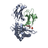


 PDBj
PDBj


 Assembly
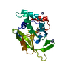


 Mass: 65.409 Da / Num. of mol.: 8 / Source method: obtained synthetically / Formula: Zn
 Mass: 60.052 Da / Num. of mol.: 1 / Source method: obtained synthetically / Formula: C2H4O2
 Mass: 18.015 Da / Num. of mol.: 179 / Source method: isolated from a natural source / Formula: H2O
 Sample preparation
 / Beamline: ID29 / Wavelength: 2.1
  Processing