 Movie
Movie Controller


+ Open data

- Basic information
| Entry | Database: PDB / ID: 6jbw | ||||||
|---|---|---|---|---|---|---|---|
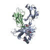
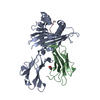
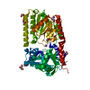
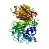
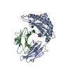
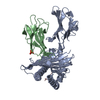
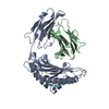
| Title | Structure of Tps1/UDP complex | ||||||
 Components Components | Trehalose-6-phosphate synthase | ||||||
 Keywords Keywords | TRANSFERASE / Trehalose-6-phosphate synthase | ||||||
| Function / homology |  Function and homology information Function and homology informationalpha,alpha-trehalose-phosphate synthase complex (UDP-forming) / alpha,alpha-trehalose-phosphate synthase (UDP-forming) / trehalose-phosphatase activity / alpha,alpha-trehalose-phosphate synthase (UDP-forming) activity / trehalose biosynthetic process / cellular response to heat / nucleotide binding / cytosol Similarity search - Function | ||||||
| Biological species |  Pyricularia oryzae 70-15 (fungus) Pyricularia oryzae 70-15 (fungus) | ||||||
| Method |  X-RAY DIFFRACTION / SYNCHROTRON / MOLECULAR REPLACEMENT / Resolution: 2.65 Å X-RAY DIFFRACTION / SYNCHROTRON / MOLECULAR REPLACEMENT / Resolution: 2.65 Å | ||||||
 Authors Authors | Wang, S. / Zhao, Y. / Wang, D. / Liu, J. | ||||||
| Funding support | 1items
| ||||||
 Citation Citation | Journal: Biochem.J. / Year: 2019 Title: Crystal structures of Magnaporthe oryzae trehalose-6-phosphate synthase (MoTps1) suggest a model for catalytic process of Tps1. Authors: Wang, S. / Zhao, Y. / Yi, L. / Shen, M. / Wang, C. / Zhang, X. / Yang, J. / Peng, Y.L. / Wang, D. / Liu, J. | ||||||
| History |
|
- Structure visualization
| Structure viewer | Molecule: MolmilJmol/JSmol |
|---|
- Downloads & links
-Download
| PDBx/mmCIF format | 6jbw.cif.gz | 195.5 KB | Display | PDBx/mmCIF format |
|---|---|---|---|---|
| PDB format | pdb6jbw.ent.gz | 151.8 KB | Display | PDB format |
| PDBx/mmJSON format | 6jbw.json.gz | Tree view | PDBx/mmJSON format | |
| Others |  Other downloads Other downloads |
-Validation report
| Arichive directory | https://data.pdbj.org/pub/pdb/validation_reports/jb/6jbwftp://data.pdbj.org/pub/pdb/validation_reports/jb/6jbw | HTTPS FTP |
|---|
-Related structure data
| Related structure data | 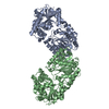 6jakC  6jbiC 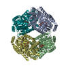 6jbrC 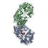 1uquS S: Starting model for refinement C: citing same article ( |
|---|---|
| Similar structure data |


-Links
 PDBj
PDBj


- Assembly
| Deposited unit | 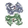
| ||||||||||||
|---|---|---|---|---|---|---|---|---|---|---|---|---|---|
| 1 | 
| ||||||||||||
| 2 | 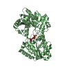
| ||||||||||||
| Unit cell |
|
-Components
| #1: Protein | Mass: 52512.719 Da / Num. of mol.: 2 Source method: isolated from a genetically manipulated source Source: (gene. exp.) Pyricularia oryzae 70-15 (fungus) / Strain: 70-15 / ATCC MYA-4617 / FGSC 8958 / Gene: MGG_03860 / Production host:  References: UniProt: G4NHF4, alpha,alpha-trehalose-phosphate synthase (UDP-forming) #2: Chemical |   Type: RNA linking / Mass: 404.161 Da / Num. of mol.: 2 / Source method: obtained synthetically / Formula: C9H14N2O12P2 / Comment: UDP*YM Type: RNA linking / Mass: 404.161 Da / Num. of mol.: 2 / Source method: obtained synthetically / Formula: C9H14N2O12P2 / Comment: UDP*YM#3: Water | ChemComp-HOH / |  Mass: 18.015 Da / Num. of mol.: 50 / Source method: isolated from a natural source / Formula: H2O Mass: 18.015 Da / Num. of mol.: 50 / Source method: isolated from a natural source / Formula: H2O |
|---|
-Experimental details
-Experiment
| Experiment | Method: X-RAY DIFFRACTION / Number of used crystals: 1 |
|---|
- Sample preparation
| Crystal | Density Matthews: 2.64 Å3/Da / Density % sol: 53.5 % |
|---|---|
| Crystal grow | Temperature: 289 K / Method: vapor diffusion, sitting drop Details: 0.2 M ammonium sulfate, 0.1 M Tris-HCl, pH 8.9, 20% PEG 3350 (v/v) |
-Data collection
| Diffraction | Mean temperature: 100 K / Serial crystal experiment: N |
|---|---|
| Diffraction source | Source: SYNCHROTRON / Site: SSRF  / Beamline: BL18U1 / Wavelength: 0.9785 Å / Beamline: BL18U1 / Wavelength: 0.9785 Å |
| Detector | Type: DECTRIS PILATUS3 6M / Detector: PIXEL / Date: May 22, 2017 |
| Radiation | Protocol: SINGLE WAVELENGTH / Monochromatic (M) / Laue (L): M / Scattering type: x-ray |
| Radiation wavelength | Wavelength: 0.9785 Å / Relative weight: 1 |
| Reflection | Resolution: 2.65→45.97 Å / Num. obs: 33303 / % possible obs: 99.7 % / Redundancy: 6.6 % / CC1/2: 1 / Rmerge(I) obs: 0.077 / Rpim(I) all: 0.023 / Net I/σ(I): 36.55 |
| Reflection shell | Resolution: 2.65→2.7 Å / Num. unique obs: 3263 / CC1/2: 0.959 |
- Processing
| Software |
| ||||||||||||||||
|---|---|---|---|---|---|---|---|---|---|---|---|---|---|---|---|---|---|
| Refinement | Method to determine structure: MOLECULAR REPLACEMENT Starting model: 1UQU Resolution: 2.65→30 Å / Cross valid method: FREE R-VALUE
| ||||||||||||||||
| Refinement step | Cycle: LAST / Resolution: 2.65→30 Å
| ||||||||||||||||
| LS refinement shell | Resolution: 2.65→2.745 Å
|
