 Movie
Movie Controller


+ Open data

- Basic information
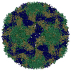
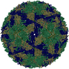
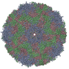
| Entry | Database: PDB / ID: 1z7s | ||||||||||||
|---|---|---|---|---|---|---|---|---|---|---|---|---|---|
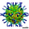
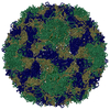
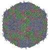
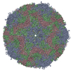
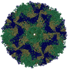
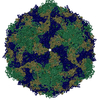
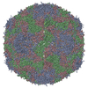
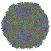
| Title | The crystal structure of coxsackievirus A21 | ||||||||||||
 Components Components | (Human COXSACKIEVIRUS ...) x 4 | ||||||||||||
 Keywords Keywords | VIRUS / PICORNAVIRUS / Coxsackievirus / A21 / Capsid Protein / Viral Protein / Icosahedral virus | ||||||||||||
| Function / homology |  Function and homology information Function and homology informationsymbiont-mediated suppression of host cytoplasmic pattern recognition receptor signaling pathway via inhibition of RIG-I activity / picornain 2A / symbiont-mediated suppression of host mRNA export from nucleus / symbiont genome entry into host cell via pore formation in plasma membrane / picornain 3C / T=pseudo3 icosahedral viral capsid / host cell cytoplasmic vesicle membrane / ribonucleoside triphosphate phosphatase activity / nucleoside-triphosphate phosphatase / channel activity ...symbiont-mediated suppression of host cytoplasmic pattern recognition receptor signaling pathway via inhibition of RIG-I activity / picornain 2A / symbiont-mediated suppression of host mRNA export from nucleus / symbiont genome entry into host cell via pore formation in plasma membrane / picornain 3C / T=pseudo3 icosahedral viral capsid / host cell cytoplasmic vesicle membrane / ribonucleoside triphosphate phosphatase activity / nucleoside-triphosphate phosphatase / channel activity / monoatomic ion transmembrane transport / DNA replication / RNA helicase activity / endocytosis involved in viral entry into host cell / symbiont-mediated activation of host autophagy / RNA-directed RNA polymerase / cysteine-type endopeptidase activity / viral RNA genome replication / RNA-directed RNA polymerase activity / virion attachment to host cell / host cell nucleus / structural molecule activity / DNA-templated transcription / proteolysis / RNA binding / zinc ion binding / ATP binding Similarity search - Function | ||||||||||||
| Biological species |  Human coxsackievirus A21 Human coxsackievirus A21 | ||||||||||||
| Method |  X-RAY DIFFRACTION / SYNCHROTRON / MOLECULAR REPLACEMENT / Resolution: 3.2 Å X-RAY DIFFRACTION / SYNCHROTRON / MOLECULAR REPLACEMENT / Resolution: 3.2 Å | ||||||||||||
 Authors Authors | Xiao, C. / Bator-Kelly, C.M. / Rieder, E. / Chipman, P.R. / Craig, A. / Kuhn, R.J. / Wimmer, E. / Rossmann, M.G. | ||||||||||||
 Citation Citation | Journal: Structure / Year: 2005 Title: The crystal structure of coxsackievirus A21 and its interaction with ICAM-1. Authors: Chuan Xiao / Carol M Bator-Kelly / Elizabeth Rieder / Paul R Chipman / Alister Craig / Richard J Kuhn / Eckard Wimmer / Michael G Rossmann /  Abstract: CVA21 and polioviruses both belong to the Enterovirus genus in the family of Picornaviridae, whereas rhinoviruses form a distinct picornavirus genus. Nevertheless, CVA21 and the major group of human ...CVA21 and polioviruses both belong to the Enterovirus genus in the family of Picornaviridae, whereas rhinoviruses form a distinct picornavirus genus. Nevertheless, CVA21 and the major group of human rhinoviruses recognize intercellular adhesion molecule-1 (ICAM-1) as their cellular receptor, whereas polioviruses use poliovirus receptor. The crystal structure of CVA21 has been determined to 3.2 A resolution. Its structure has greater similarity to poliovirus structures than to other known picornavirus structures. Cryo-electron microscopy (cryo-EM) was used to determine an 8.0 A resolution structure of CVA21 complexed with an ICAM-1 variant, ICAM-1(Kilifi). The cryo-EM map was fitted with the crystal structures of ICAM-1 and CVA21. Significant differences in the structure of CVA21 with respect to the poliovirus structures account for the inability of ICAM-1 to bind polioviruses. The interface between CVA21 and ICAM-1 has shape and electrostatic complementarity with many residues being conserved among those CVAs that bind ICAM-1. | ||||||||||||
| History |
| ||||||||||||
| Remark 600 | HETEROGEN THE N-TERMINAL OF VP4 (CHAIN 4) IS MYRISTYLATED WHICH MEANS THE MYR IS COVALENTLY LINKED ... HETEROGEN THE N-TERMINAL OF VP4 (CHAIN 4) IS MYRISTYLATED WHICH MEANS THE MYR IS COVALENTLY LINKED TO THE GLY OF VP4. THEREFORE, THE O2 ATOM ON MYR IS EXPECTED TO BE MISSING AFTER MYRISTYLATED. | ||||||||||||
| Remark 999 | SEQUENCE THE COMPLETE GENOME SEQUENCE FOR HUMAN COXSACKIEVIRUS A21 IS AVAILABLE AT GENBANK WITH ...SEQUENCE THE COMPLETE GENOME SEQUENCE FOR HUMAN COXSACKIEVIRUS A21 IS AVAILABLE AT GENBANK WITH ACCESSION CODE AF546702 (GI:3304568). THE INDEX OF THIS VIRUS IS 00.052.0.01.005.01.021 WHICH IS AVAILABLE AT ICTVDB: HTTP://WWW.NCBI.NLM.NIH.GOV/ICTVDB/ICTV/INDEX.HTM. |
- Structure visualization
| Structure viewer | Molecule: MolmilJmol/JSmol |
|---|
- Downloads & links
-Download
| PDBx/mmCIF format | 1z7s.cif.gz | 184.9 KB | Display | PDBx/mmCIF format |
|---|---|---|---|---|
| PDB format | pdb1z7s.ent.gz | 144.1 KB | Display | PDB format |
| PDBx/mmJSON format | 1z7s.json.gz | Tree view | PDBx/mmJSON format | |
| Others |  Other downloads Other downloads |
-Validation report
| Arichive directory | https://data.pdbj.org/pub/pdb/validation_reports/z7/1z7sftp://data.pdbj.org/pub/pdb/validation_reports/z7/1z7s | HTTPS FTP |
|---|
-Related structure data


-Links
 PDBj
PDBj


- Assembly
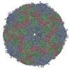
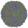
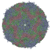
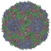
| Deposited unit | 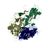
| ||||||||||||||||||
|---|---|---|---|---|---|---|---|---|---|---|---|---|---|---|---|---|---|---|---|
| 1 | x 60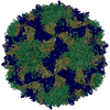
| ||||||||||||||||||
| 2 |
| ||||||||||||||||||
| 3 | x 5
| ||||||||||||||||||
| 4 | x 6
| ||||||||||||||||||
| 5 | 
| ||||||||||||||||||
| 6 | x 5
| ||||||||||||||||||
| Unit cell |
| ||||||||||||||||||
| Symmetry | Point symmetry: (Hermann–Mauguin notation: 532 / Schoenflies symbol: I (icosahedral)) | ||||||||||||||||||
| Noncrystallographic symmetry (NCS) | NCS oper:
|
-Components
-Human COXSACKIEVIRUS ... , 4 types, 4 molecules 1234
| #1: Protein | Mass: 33264.262 Da / Num. of mol.: 1 / Fragment: Viral Protein 1 / Source method: isolated from a natural source / Details: the nature host of this virus is human / Source: (natural) Human coxsackievirus A21 / Genus: Enterovirus / Cell line: Hela / Species: Human enterovirus C / Strain: Kuykendall / References: UniProt: Q71LY2, UniProt: P22055*PLUS |
|---|---|
| #2: Protein | Mass: 29918.537 Da / Num. of mol.: 1 / Fragment: Viral Protein 2 / Source method: isolated from a natural source / Details: the nature host of this virus is human / Source: (natural) Human coxsackievirus A21 / Genus: Enterovirus / Cell line: Hela / Species: Human enterovirus C / Strain: Kuykendall / References: UniProt: Q71LY2, UniProt: P22055*PLUS |
| #3: Protein | Mass: 26568.689 Da / Num. of mol.: 1 / Fragment: Viral Protein 3 / Source method: isolated from a natural source / Details: the nature host of this virus is human / Source: (natural) Human coxsackievirus A21 / Genus: Enterovirus / Cell line: Hela / Species: Human enterovirus C / Strain: Kuykendall / References: UniProt: Q71LY2, UniProt: P22055*PLUS |
| #4: Protein | Mass: 7310.922 Da / Num. of mol.: 1 / Fragment: Viral Protein 4 / Source method: isolated from a natural source / Details: the nature host of this virus is human / Source: (natural) Human coxsackievirus A21 / Genus: Enterovirus / Cell line: Hela / Species: Human enterovirus C / Strain: Kuykendall / References: UniProt: Q71LY2, UniProt: P22055*PLUS |
-Non-polymers , 4 types, 57 molecules 


| #5: Chemical | ChemComp-CA /  Mass: 40.078 Da / Num. of mol.: 1 / Source method: obtained synthetically / Formula: Ca Mass: 40.078 Da / Num. of mol.: 1 / Source method: obtained synthetically / Formula: Ca | ||||
|---|---|---|---|---|---|
| #6: Chemical |  Mass: 228.371 Da / Num. of mol.: 2 / Source method: obtained synthetically / Formula: C14H28O2 Mass: 228.371 Da / Num. of mol.: 2 / Source method: obtained synthetically / Formula: C14H28O2#7: Chemical | ChemComp-5GP / |  Mass: 363.221 Da / Num. of mol.: 1 / Source method: obtained synthetically / Formula: C10H14N5O8P Mass: 363.221 Da / Num. of mol.: 1 / Source method: obtained synthetically / Formula: C10H14N5O8P#8: Water | ChemComp-HOH / | Mass: 18.015 Da / Num. of mol.: 53 / Source method: isolated from a natural source / Formula: H2O |
-Details
| Has protein modification | Y |
|---|
-Experimental details
-Experiment
| Experiment | Method: X-RAY DIFFRACTION / Number of used crystals: 2 |
|---|
- Sample preparation
| Crystal |
| |||||||||||||||
|---|---|---|---|---|---|---|---|---|---|---|---|---|---|---|---|---|
| Crystal grow |
|
-Data collection
| Diffraction | Mean temperature: 100 K |
|---|---|
| Diffraction source | Source: SYNCHROTRON / Site: APS / Beamline: 14-BM-C / Wavelength: 1 Å |
| Detector | Type: ADSC QUANTUM 4 / Detector: CCD / Date: Jan 1, 2000 |
| Radiation | Protocol: SINGLE WAVELENGTH / Monochromatic (M) / Laue (L): M / Scattering type: x-ray |
| Radiation wavelength | Wavelength: 1 Å / Relative weight: 1 |
| Reflection | Resolution: 3.2→50 Å / Num. all: 110120 / Num. obs: 106927 / % possible obs: 97.1 % / Observed criterion σ(F): 3 / Observed criterion σ(I): 3 / Redundancy: 17 % / Rmerge(I) obs: 0.119 |
| Reflection shell | Resolution: 3.2→3.31 Å / Redundancy: 11 % / Rmerge(I) obs: 0.238 / % possible all: 88.9 |
- Processing
| Software |
| ||||||||||||||||||||||||||||||||||||
|---|---|---|---|---|---|---|---|---|---|---|---|---|---|---|---|---|---|---|---|---|---|---|---|---|---|---|---|---|---|---|---|---|---|---|---|---|---|
| Refinement | Method to determine structure: MOLECULAR REPLACEMENT / Resolution: 3.2→49.72 Å / Rfactor Rfree error: 0.002 / Data cutoff high absF: 17050733.6 / Data cutoff low absF: 0 / Isotropic thermal model: RESTRAINED / Cross valid method: THROUGHOUT / σ(F): 0
| ||||||||||||||||||||||||||||||||||||
| Solvent computation | Solvent model: FLAT MODEL / Bsol: 30.6012 Å2 / ksol: 3.60356 e/Å3 | ||||||||||||||||||||||||||||||||||||
| Displacement parameters | Biso mean: 51.2 Å2
| ||||||||||||||||||||||||||||||||||||
| Refine analyze |
| ||||||||||||||||||||||||||||||||||||
| Refinement step | Cycle: LAST / Resolution: 3.2→49.72 Å
| ||||||||||||||||||||||||||||||||||||
| Refine LS restraints |
| ||||||||||||||||||||||||||||||||||||
| Refine LS restraints NCS | NCS model details: CONSTR | ||||||||||||||||||||||||||||||||||||
| LS refinement shell | Resolution: 3.2→3.4 Å / Rfactor Rfree error: 0.008 / Total num. of bins used: 6
| ||||||||||||||||||||||||||||||||||||
| Xplor file |
|
