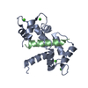
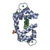
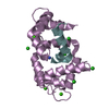
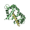
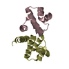
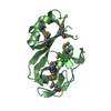
Entry Database : PDB / ID : 1yr5Title 1.7-A structure of calmodulin bound to a peptide from DAP kinase 19-mer from Death-associated protein kinase 1 calmodulin Keywords / / Function / homology Function Domain/homology Component
/ / / / / / / / / / / / / / / / / / / / / / / / / / / / / / / / / / / / / / / / / / / / / / / / / / / / / / / / / / / / / / / / / / / / / / / / / / / / / / / / / / / / / / / / / / / / / / / / / / / / / / / / / / / / / / / / / / / / / / / / / / / / / / / / / / / / / / / / / / / / / / / / / / / / / / / / / / Biological species Homo sapiens (human)Method / / / Resolution : 1.7 Å Authors Kursula, P. / Vahokoski, J. / Wilmanns, M. Journal : To be Published Title : Recognition of human death-associated protein kinases by calmodulinAuthors : Kursula, P. / Vahokoski, J. / Wilmanns, M. History Deposition Feb 3, 2005 Deposition site / Processing site Revision 1.0 Jul 4, 2006 Provider / Type Revision 1.1 Apr 30, 2008 Group Revision 1.2 Jul 13, 2011 Group / Version format complianceRevision 1.3 Nov 20, 2019 Group / Derived calculationsCategory / pdbx_struct_conn_angle / struct_connItem Revision 1.4 Oct 25, 2023 Group Data collection / Database references ... Data collection / Database references / Derived calculations / Refinement description Category chem_comp_atom / chem_comp_bond ... chem_comp_atom / chem_comp_bond / database_2 / pdbx_initial_refinement_model / struct_site Item _database_2.pdbx_DOI / _database_2.pdbx_database_accession ... _database_2.pdbx_DOI / _database_2.pdbx_database_accession / _struct_site.pdbx_auth_asym_id / _struct_site.pdbx_auth_comp_id / _struct_site.pdbx_auth_seq_id
Show all Show less
 Movie
Movie Controller


 Open data
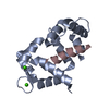
 Basic information
 Components
 Keywords
 Function and homology information
 Homo sapiens (human)
 X-RAY DIFFRACTION /
  Authors
 Citation
 Structure visualization
 Downloads & links
 Other downloads

 PDBj
PDBj


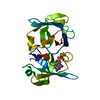
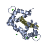
 Assembly
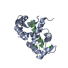


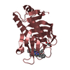
 Mass: 40.078 Da / Num. of mol.: 4 / Source method: obtained synthetically / Formula: Ca
 Mass: 18.015 Da / Num. of mol.: 144 / Source method: isolated from a natural source / Formula: H2O
 Sample preparation
 / Beamline: BW7A / Wavelength: 0.938 Å
 Processing