[English] 日本語
 Yorodumi
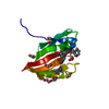
- PDB-1yr1: Structure of the major extracytoplasmic domain of the trans isome... -
+ Open data
- Basic information
| Entry | Database: PDB / ID: 1yr1 | ||||||
|---|---|---|---|---|---|---|---|
| Title | Structure of the major extracytoplasmic domain of the trans isomer of the bacterial cell division protein divib from geobacillus stearothermophilus | ||||||
 Components Components | cell-division initiation protein | ||||||
 Keywords Keywords | CELL CYCLE / cell-division initiation protein / DIVIB / FTSQ / DIVISOME | ||||||
| Function / homology |  Function and homology information Function and homology information | ||||||
| Biological species |   Geobacillus stearothermophilus (bacteria) Geobacillus stearothermophilus (bacteria) | ||||||
| Method | SOLUTION NMR / torsion angle dynamics (CANDID, CYANA), simulated annealing (XPLOR) | ||||||
 Authors Authors | Robson, S.A. / King, G.F. | ||||||
 Citation Citation |  Journal: Proc.Natl.Acad.Sci.USA / Year: 2006 Journal: Proc.Natl.Acad.Sci.USA / Year: 2006Title: Domain architecture and structure of the bacterial cell division protein DivIB. Authors: Robson, S.A. / King, G.F. #1: Journal: J.Bacteriol. / Year: 1989 Title: Cloning and expression of a Bacillus subtilis division initiation gene for which a homolog has not been identified in another organism Authors: Harry, E.J. / Wake, R.G. #2: Journal: J.Bacteriol. / Year: 1989 Title: Nucleotide sequence and insertional inactivation of a Bacillus subtilis gene that affects cell division, sporulation, and temperature sensitivity. Authors: Beall, B. / Lutkenhaus, J. #3:  Journal: Mol.Microbiol. / Year: 1997 Journal: Mol.Microbiol. / Year: 1997Title: DivIB, FtsZ and cell division in Bacillus subtilis. Authors: Rowland, S.L. / Katis, V.L. / Partridge, S.R. / Wake, R.G. #4:  Journal: J.Bacteriol. / Year: 1999 Journal: J.Bacteriol. / Year: 1999Title: Membrane-bound division proteins DivIB and DivIC of Bacillus subtilis function solely through their external domains in both vegetative and sporulation division. Authors: Katis, V.L. / Wake, R.G. | ||||||
| History |
| ||||||
| Remark 650 | HELIX DETERMINATION METHOD: AUTHOR DETERMINED | ||||||
| Remark 999 | SEQUENCE DivIB fragment was subcloned as a translational fusion to the C-terminus of Schistosoma ...SEQUENCE DivIB fragment was subcloned as a translational fusion to the C-terminus of Schistosoma japonicum glutathione S-transferase. The gene sequence encoding this protein domain was cloned using chromosomal DNA from Geobacillus stearothermophilus. The sequence matches that of the corresponding domain of the DivIB ortholog from Geobacillus kaustophilus HTA426 (SWISS-PROT entry Q5L0X5_GEOKA). |
- Structure visualization
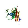
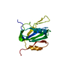
| Structure viewer | Molecule:  Molmil Molmil Jmol/JSmol Jmol/JSmol |
|---|
- Downloads & links
- Download
| PDBx/mmCIF format |  1yr1.cif.gz 1yr1.cif.gz | 902.2 KB | Display |  PDBx/mmCIF format PDBx/mmCIF format |
|---|---|---|---|---|
| PDB format |  pdb1yr1.ent.gz pdb1yr1.ent.gz | 758 KB | Display |  PDB format PDB format |
| PDBx/mmJSON format |  1yr1.json.gz 1yr1.json.gz | Tree view |  PDBx/mmJSON format PDBx/mmJSON format | |
| Others |  Other downloads Other downloads |
-Validation report
| Summary document |  1yr1_validation.pdf.gz 1yr1_validation.pdf.gz | 345.5 KB | Display |  wwPDB validaton report wwPDB validaton report |
|---|---|---|---|---|
| Full document |  1yr1_full_validation.pdf.gz 1yr1_full_validation.pdf.gz | 518.1 KB | Display | |
| Data in XML |  1yr1_validation.xml.gz 1yr1_validation.xml.gz | 51 KB | Display | |
| Data in CIF |  1yr1_validation.cif.gz 1yr1_validation.cif.gz | 84.4 KB | Display | |
| Arichive directory |  https://data.pdbj.org/pub/pdb/validation_reports/yr/1yr1 https://data.pdbj.org/pub/pdb/validation_reports/yr/1yr1 ftp://data.pdbj.org/pub/pdb/validation_reports/yr/1yr1 ftp://data.pdbj.org/pub/pdb/validation_reports/yr/1yr1 | HTTPS FTP |
-Related structure data
- Links
Links
- Assembly
| Deposited unit | 
| |||||||||
|---|---|---|---|---|---|---|---|---|---|---|
| 1 |
| |||||||||
| NMR ensembles |
|
- Components
| #1: Protein | Mass: 13365.084 Da / Num. of mol.: 1 Source method: isolated from a genetically manipulated source Source: (gene. exp.)   Geobacillus stearothermophilus (bacteria) Geobacillus stearothermophilus (bacteria)Gene: divIB / Plasmid: pSAR19 / Species (production host): Escherichia coli / Production host:  |
|---|
-Experimental details
-Experiment
| Experiment | Method: SOLUTION NMR | ||||||||||||||||||||
|---|---|---|---|---|---|---|---|---|---|---|---|---|---|---|---|---|---|---|---|---|---|
| NMR experiment |
|
- Sample preparation
| Details |
| |||||||||
|---|---|---|---|---|---|---|---|---|---|---|
| Sample conditions | Ionic strength: 0.16 / pH: 6 / Pressure: ambient / Temperature: 308 K |
-NMR measurement
| Radiation | Protocol: SINGLE WAVELENGTH / Monochromatic (M) / Laue (L): M / Scattering type: x-ray | |||||||||||||||
|---|---|---|---|---|---|---|---|---|---|---|---|---|---|---|---|---|
| Radiation wavelength | Relative weight: 1 | |||||||||||||||
| NMR spectrometer |
|
- Processing
| NMR software |
| ||||||||||||||||||||||||
|---|---|---|---|---|---|---|---|---|---|---|---|---|---|---|---|---|---|---|---|---|---|---|---|---|---|
| Refinement | Method: torsion angle dynamics (CANDID, CYANA), simulated annealing (XPLOR) Software ordinal: 1 Details: The structures are based on a total of 2709 NOE-derived distance restraints, 82 restraints defining 41 hydrogen bonds, and 196 dihedral angle restraints | ||||||||||||||||||||||||
| NMR representative | Selection criteria: lowest energy | ||||||||||||||||||||||||
| NMR ensemble | Conformer selection criteria: lowest energy / Conformers calculated total number: 60 / Conformers submitted total number: 25 |
 Movie
Movie Controller




 PDBj
PDBj NMRPipe