 Movie
Movie Controller

[English] 日本語
 Yorodumi
- PDB-1jsg: CRYSTAL STRUCTURE OF P14TCL1, AN ONCOGENE PRODUCT INVOLVED IN T-C... -
+ Open data

- Basic information
| Entry | Database: PDB / ID: 1jsg | ||||||
|---|---|---|---|---|---|---|---|
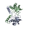
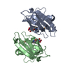
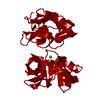
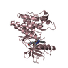
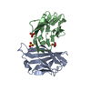
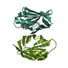
| Title | CRYSTAL STRUCTURE OF P14TCL1, AN ONCOGENE PRODUCT INVOLVED IN T-CELL PROLYMPHOCYTIC LEUKEMIA, REVEALS A NOVEL B-BARREL TOPOLOGY | ||||||
 Components Components | ONCOGENE PRODUCT P14TCL1 | ||||||
 Keywords Keywords | PROTO-ONCOGENE / CL1 GENE / T CELL LEUKEMIA / ONCOPROTEIN / MICROSOME | ||||||
| Function / homology |  Function and homology information Function and homology informationprotein serine/threonine kinase activator activity / intracellular signal transduction / protein kinase binding / endoplasmic reticulum / nucleoplasm / identical protein binding / nucleus / cytosol Similarity search - Function | ||||||
| Biological species |  Homo sapiens (human) Homo sapiens (human) | ||||||
| Method |  X-RAY DIFFRACTION / Resolution: 2.5 Å X-RAY DIFFRACTION / Resolution: 2.5 Å | ||||||
 Authors Authors | Hoh, F. / Yang, Y.-S. / Guignard, L. / Padilla, A. / Stern, R.-H. / Lhoste, J.-M. / Van Tilbeurgh, H. | ||||||
 Citation Citation | Journal: Structure / Year: 1998 Title: Crystal structure of p14TCL1, an oncogene product involved in T-cell prolymphocytic leukemia, reveals a novel beta-barrel topology. Authors: Hoh, F. / Yang, Y.S. / Guignard, L. / Padilla, A. / Stern, M.H. / Lhoste, J.M. / van Tilbeurgh, H. | ||||||
| History |
|
- Structure visualization
| Structure viewer | Molecule: MolmilJmol/JSmol |
|---|
- Downloads & links
-Download
| PDBx/mmCIF format | 1jsg.cif.gz | 34.6 KB | Display | PDBx/mmCIF format |
|---|---|---|---|---|
| PDB format | pdb1jsg.ent.gz | 23.9 KB | Display | PDB format |
| PDBx/mmJSON format | 1jsg.json.gz | Tree view | PDBx/mmJSON format | |
| Others |  Other downloads Other downloads |
-Validation report
| Arichive directory | https://data.pdbj.org/pub/pdb/validation_reports/js/1jsgftp://data.pdbj.org/pub/pdb/validation_reports/js/1jsg | HTTPS FTP |
|---|
-Related structure data
| Similar structure data |
|---|


-Links
 PDBj
PDBj- Assembly
| Deposited unit | 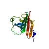
| ||||||||
|---|---|---|---|---|---|---|---|---|---|
| 1 | 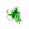
| ||||||||
| Unit cell |
| ||||||||
| Details | P14 IS INVOLVED IN RARE FORMS OF LEUKEMIA BUT ITS CELLULAR ROLE REMAINS UNKNOWN. THERE EXISTS EVIDENCE FOR DIMER FORMATION IN SOLUTION BUT THE PROTEIN IS PRESENT AS A MONOMER IN THE ASYMMETRIC UNIT. A TIGHT DIMER CONTACT IS GENERATED BY THE TWO-FOLD CRYSTAL C-AXIS. |
-Components
| #1: Protein | Mass: 13475.537 Da / Num. of mol.: 1 / Source method: isolated from a natural source / Source: (natural) Homo sapiens (human) / Cell: T-CELL / References: UniProt: P56279 |
|---|---|
| #2: Water | ChemComp-HOH /  Mass: 18.015 Da / Num. of mol.: 49 / Source method: isolated from a natural source / Formula: H2O Mass: 18.015 Da / Num. of mol.: 49 / Source method: isolated from a natural source / Formula: H2O |
-Experimental details
-Experiment
| Experiment | Method: X-RAY DIFFRACTION |
|---|
- Sample preparation
| Crystal | Density Matthews: 3.52 Å3/Da / Density % sol: 65.02 % | ||||||||||||||||||||||||
|---|---|---|---|---|---|---|---|---|---|---|---|---|---|---|---|---|---|---|---|---|---|---|---|---|---|
| Crystal grow | *PLUS Method: vapor diffusion, hanging drop | ||||||||||||||||||||||||
| Components of the solutions | *PLUS
|
-Data collection
| Radiation | Scattering type: x-ray |
|---|---|
| Radiation wavelength | Relative weight: 1 |
| Reflection | Biso Wilson estimate: 37 Å2 |
| Reflection | *PLUS Highest resolution: 2.5 Å / Lowest resolution: 40 Å / Num. obs: 6870 / % possible obs: 99.4 % / Redundancy: 8.8 % / Num. measured all: 60407 / Rmerge(I) obs: 0.099 |
| Reflection shell | *PLUS Highest resolution: 2.5 Å / Lowest resolution: 2.6 Å / % possible obs: 99.9 % / Rmerge(I) obs: 0.288 |
- Processing
| Software |
| ||||||||||||||||||||||||||||||||||||||||||||||||||||||||||||
|---|---|---|---|---|---|---|---|---|---|---|---|---|---|---|---|---|---|---|---|---|---|---|---|---|---|---|---|---|---|---|---|---|---|---|---|---|---|---|---|---|---|---|---|---|---|---|---|---|---|---|---|---|---|---|---|---|---|---|---|---|---|
| Refinement | Resolution: 2.5→8 Å / Rfactor Rfree error: 0.01 / Data cutoff high absF: 10000000 / Data cutoff low absF: 0.001 / Isotropic thermal model: GROUP / Cross valid method: THROUGHOUT / σ(F): 0
| ||||||||||||||||||||||||||||||||||||||||||||||||||||||||||||
| Displacement parameters | Biso mean: 29.6 Å2 | ||||||||||||||||||||||||||||||||||||||||||||||||||||||||||||
| Refine analyze |
| ||||||||||||||||||||||||||||||||||||||||||||||||||||||||||||
| Refinement step | Cycle: LAST / Resolution: 2.5→8 Å
| ||||||||||||||||||||||||||||||||||||||||||||||||||||||||||||
| Refine LS restraints |
| ||||||||||||||||||||||||||||||||||||||||||||||||||||||||||||
| LS refinement shell | Resolution: 2.5→2.65 Å / Rfactor Rfree error: 0.034 / Total num. of bins used: 6
| ||||||||||||||||||||||||||||||||||||||||||||||||||||||||||||
| Xplor file |
| ||||||||||||||||||||||||||||||||||||||||||||||||||||||||||||
| Software | *PLUS Version: 3.851 / Classification: refinement | ||||||||||||||||||||||||||||||||||||||||||||||||||||||||||||
| Refine LS restraints | *PLUS
|
