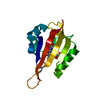
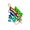
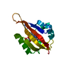
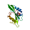
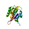
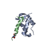
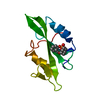
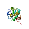
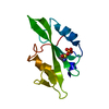
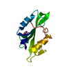
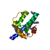
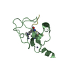
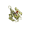
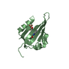
SIGNALING PROTEIN / flavoprotein / LOV / Blue light photoreceptor
Function / homology
Function and homology information
chloroplast relocation / phototropism / blue light signaling pathway / stomatal movement / blue light photoreceptor activity / response to blue light / plastid / circadian rhythm / kinase activity / FMN binding ...chloroplast relocation / phototropism / blue light signaling pathway / stomatal movement / blue light photoreceptor activity / response to blue light / plastid / circadian rhythm / kinase activity / FMN binding / protein autophosphorylation / non-specific serine/threonine protein kinase / protein serine kinase activity / protein serine/threonine kinase activity / Golgi apparatus / ATP binding / membrane / identical protein binding / nucleus / plasma membrane / cytoplasm Similarity search - Function
PAS domain / PAS-associated, C-terminal / PAC domain profile. / PAC motif / Motif C-terminal to PAS motifs (likely to contribute to PAS structural domain) / PAS domain / Beta-Lactamase / PAS domain / PAS repeat profile. / PAS domain ...PAS domain / PAS-associated, C-terminal / PAC domain profile. / PAC motif / Motif C-terminal to PAS motifs (likely to contribute to PAS structural domain) / PAS domain / Beta-Lactamase / PAS domain / PAS repeat profile. / PAS domain / PAS domain superfamily / Serine/threonine-protein kinase, active site / Serine/Threonine protein kinases active-site signature. / Protein kinase domain / Serine/Threonine protein kinases, catalytic domain / Protein kinase, ATP binding site / Protein kinases ATP-binding region signature. / Protein kinase domain profile. / Protein kinase domain / Protein kinase-like domain superfamily / 2-Layer Sandwich / Alpha Beta Similarity search - Domain/homology
Phototropin-2 / Defective in chloroplast avoidance protein 1 / Non-phototropic hypocotyl 1-like protein 1 / AtKin7 ...Defective in chloroplast avoidance protein 1 / Non-phototropic hypocotyl 1-like protein 1 / AtKin7 / NPH1-like protein 1
Mass: 13278.843 Da / Num. of mol.: 2 / Fragment: LOV DOMAIN (UNP Residues 385-496) Source method: isolated from a genetically manipulated source Source: (gene. exp.) Arabidopsis thaliana (thale cress) / Gene: PHOT2, CAV1, KIN7, NPL1, At5g58140, K21L19.6 / Production host: Escherichia coli (E. coli) References: UniProt: P93025, non-specific serine/threonine protein kinase
Resolution: 1.2→50 Å / Cor.coef. Fo:Fc: 0.965 / Cor.coef. Fo:Fc free: 0.963 / SU B: 0.646 / SU ML: 0.03 / Cross valid method: THROUGHOUT / ESU R: 0.046 / ESU R Free: 0.045 / Stereochemistry target values: MAXIMUM LIKELIHOOD / Details: HYDROGENS HAVE BEEN ADDED IN THE RIDING POSITIONS
Rfactor
Num. reflection
% reflection
Selection details
Rfree
0.21146
3648
5 %
RANDOM
Rwork
0.2011
-
-
-
obs
0.20162
68718
99.74 %
-
Solvent computation
Ion probe radii: 0.8 Å / Shrinkage radii: 0.8 Å / VDW probe radii: 1.2 Å / Solvent model: BABINET MODEL WITH MASK
Displacement parameters
Biso mean: 18.692 Å2
Baniso -1
Baniso -2
Baniso -3
1-
0.56 Å2
0 Å2
-0 Å2
2-
-
0.56 Å2
-0 Å2
3-
-
-
-1.13 Å2
Refinement step
Cycle: LAST / Resolution: 1.2→50 Å
Protein
Nucleic acid
Ligand
Solvent
Total
Num. atoms
1788
0
62
220
2070
Refine LS restraints
Refine-ID
Type
Dev ideal
Dev ideal target
Number
X-RAY DIFFRACTION
r_bond_refined_d
0.007
0.022
1906
X-RAY DIFFRACTION
r_bond_other_d
X-RAY DIFFRACTION
r_angle_refined_deg
1.157
1.999
2588
X-RAY DIFFRACTION
r_angle_other_deg
X-RAY DIFFRACTION
r_dihedral_angle_1_deg
6.101
5
220
X-RAY DIFFRACTION
r_dihedral_angle_2_deg
26.987
24.231
104
X-RAY DIFFRACTION
r_dihedral_angle_3_deg
10.828
15
333
X-RAY DIFFRACTION
r_dihedral_angle_4_deg
17.574
15
18
X-RAY DIFFRACTION
r_chiral_restr
0.08
0.2
276
X-RAY DIFFRACTION
r_gen_planes_refined
0.006
0.021
1465
X-RAY DIFFRACTION
r_gen_planes_other
X-RAY DIFFRACTION
r_nbd_refined
X-RAY DIFFRACTION
r_nbd_other
X-RAY DIFFRACTION
r_nbtor_refined
X-RAY DIFFRACTION
r_nbtor_other
X-RAY DIFFRACTION
r_xyhbond_nbd_refined
X-RAY DIFFRACTION
r_xyhbond_nbd_other
X-RAY DIFFRACTION
r_metal_ion_refined
X-RAY DIFFRACTION
r_metal_ion_other
X-RAY DIFFRACTION
r_symmetry_vdw_refined
X-RAY DIFFRACTION
r_symmetry_vdw_other
X-RAY DIFFRACTION
r_symmetry_hbond_refined
X-RAY DIFFRACTION
r_symmetry_hbond_other
X-RAY DIFFRACTION
r_symmetry_metal_ion_refined
X-RAY DIFFRACTION
r_symmetry_metal_ion_other
X-RAY DIFFRACTION
r_mcbond_it
0.657
1.5
1090
X-RAY DIFFRACTION
r_mcbond_other
X-RAY DIFFRACTION
r_mcangle_it
1.261
2
1772
X-RAY DIFFRACTION
r_scbond_it
1.723
3
816
X-RAY DIFFRACTION
r_scangle_it
2.839
4.5
814
X-RAY DIFFRACTION
r_rigid_bond_restr
X-RAY DIFFRACTION
r_sphericity_free
X-RAY DIFFRACTION
r_sphericity_bonded
LS refinement shell
Resolution: 1.2→1.231 Å / Total num. of bins used: 20
Rfactor
Num. reflection
% reflection
Rfree
0.305
235
-
Rwork
0.314
4864
-
obs
-
-
96.96 %
+
About Yorodumi
-
News
-
Feb 9, 2022. New format data for meta-information of EMDB entries
New format data for meta-information of EMDB entries
Version 3 of the EMDB header file is now the official format.
The previous official version 1.9 will be removed from the archive.
In the structure databanks used in Yorodumi, some data are registered as the other names, "COVID-19 virus" and "2019-nCoV". Here are the details of the virus and the list of structure data.
Jan 31, 2019. EMDB accession codes are about to change! (news from PDBe EMDB page)
EMDB accession codes are about to change! (news from PDBe EMDB page)
The allocation of 4 digits for EMDB accession codes will soon come to an end. Whilst these codes will remain in use, new EMDB accession codes will include an additional digit and will expand incrementally as the available range of codes is exhausted. The current 4-digit format prefixed with “EMD-” (i.e. EMD-XXXX) will advance to a 5-digit format (i.e. EMD-XXXXX), and so on. It is currently estimated that the 4-digit codes will be depleted around Spring 2019, at which point the 5-digit format will come into force.
The EM Navigator/Yorodumi systems omit the EMD- prefix.
Related info.:Q: What is EMD? / ID/Accession-code notation in Yorodumi/EM Navigator
Yorodumi is a browser for structure data from EMDB, PDB, SASBDB, etc.
This page is also the successor to EM Navigator detail page, and also detail information page/front-end page for Omokage search.
The word "yorodu" (or yorozu) is an old Japanese word meaning "ten thousand". "mi" (miru) is to see.
Related info.:EMDB / PDB / SASBDB / Comparison of 3 databanks / Yorodumi Search / Aug 31, 2016. New EM Navigator & Yorodumi / Yorodumi Papers / Jmol/JSmol / Function and homology information / Changes in new EM Navigator and Yorodumi
 Movie
Movie Controller


 Open data
 Basic information
 Components
 Keywords
 Function and homology information
 X-RAY DIFFRACTION /
  Authors
 Citation
 Structure visualization
 Downloads & links
 Other downloads
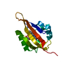

 PDBj
PDBj


 Assembly


 Mass: 456.344 Da / Num. of mol.: 2 / Source method: obtained synthetically / Formula: C17H21N4O9P
 Mass: 18.015 Da / Num. of mol.: 220 / Source method: isolated from a natural source / Formula: H2O
 Sample preparation
 / Beamline: 12.3.1 / Wavelength: 1.115922 Å
 Processing