 Movie
Movie Controller

[English] 日本語
 Yorodumi
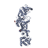
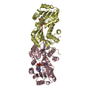
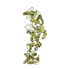
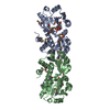
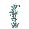
- PDB-1snd: STAPHYLOCOCCAL NUCLEASE DIMER CONTAINING A DELETION OF RESIDUES 1... -
+ Open data

- Basic information
| Entry | Database: PDB / ID: 1snd | ||||||
|---|---|---|---|---|---|---|---|
| Title | STAPHYLOCOCCAL NUCLEASE DIMER CONTAINING A DELETION OF RESIDUES 114-119 COMPLEXED WITH CALCIUM CHLORIDE AND THE COMPETITIVE INHIBITOR DEOXYTHYMIDINE-3',5'-DIPHOSPHATE | ||||||
 Components Components | STAPHYLOCOCCAL NUCLEASE DIMER | ||||||
 Keywords Keywords | HYDROLASE / NUCLEASE / ENDONUCLEASE | ||||||
| Function / homology |  Function and homology information Function and homology informationmicrococcal nuclease / 3' overhang single-stranded DNA endonuclease activity / nucleic acid binding / extracellular region / membrane / metal ion binding Similarity search - Function | ||||||
| Biological species |   Staphylococcus aureus (bacteria) Staphylococcus aureus (bacteria) | ||||||
| Method |  X-RAY DIFFRACTION / Resolution: 1.84 Å X-RAY DIFFRACTION / Resolution: 1.84 Å | ||||||
 Authors Authors | Green, S.M. / Gittis, A.G. / Meeker, A.K. / Lattman, E.E. | ||||||
 Citation Citation | Journal: Nat.Struct.Biol. / Year: 1995 Title: One-step evolution of a dimer from a monomeric protein. Authors: Green, S.M. / Gittis, A.G. / Meeker, A.K. / Lattman, E.E. | ||||||
| History |
|
- Structure visualization
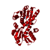
| Structure viewer | Molecule: MolmilJmol/JSmol |
|---|
- Downloads & links
-Download
| PDBx/mmCIF format | 1snd.cif.gz | 64.9 KB | Display | PDBx/mmCIF format |
|---|---|---|---|---|
| PDB format | pdb1snd.ent.gz | 48.9 KB | Display | PDB format |
| PDBx/mmJSON format | 1snd.json.gz | Tree view | PDBx/mmJSON format | |
| Others |  Other downloads Other downloads |
-Validation report
| Arichive directory | https://data.pdbj.org/pub/pdb/validation_reports/sn/1sndftp://data.pdbj.org/pub/pdb/validation_reports/sn/1snd | HTTPS FTP |
|---|
-Related structure data
| Similar structure data |
|---|


-Links
 PDBj
PDBj- Assembly
| Deposited unit | 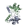
| |||||||||
|---|---|---|---|---|---|---|---|---|---|---|
| 1 | 
| |||||||||
| 2 |
| |||||||||
| Unit cell |
| |||||||||
| Components on special symmetry positions |
|
-Components
| #1: Protein | Mass: 16126.520 Da / Num. of mol.: 2 / Mutation: DEL(114-119) Source method: isolated from a genetically manipulated source Details: ALTHOUGH THE LIGANDS WERE ADDED FOR CRYSTALLIZATION, THEY WERE NOT OBSERVED IN THE ELECTRON DENSITY MAPS AND WERE NOT INCLUDED IN THE FINAL STRUCTURE Source: (gene. exp.) Staphylococcus aureus (bacteria) / Strain: FOGGI / Plasmid: LAMBDA PL12 / Production host: #2: Water | ChemComp-HOH / |  Mass: 18.015 Da / Num. of mol.: 138 / Source method: isolated from a natural source / Formula: H2O Mass: 18.015 Da / Num. of mol.: 138 / Source method: isolated from a natural source / Formula: H2O |
|---|
-Experimental details
-Experiment
| Experiment | Method: X-RAY DIFFRACTION |
|---|
- Sample preparation
| Crystal | Density Matthews: 3.72 Å3/Da / Density % sol: 66.9 % | ||||||||||||||||||||||||||||||||||||||||||
|---|---|---|---|---|---|---|---|---|---|---|---|---|---|---|---|---|---|---|---|---|---|---|---|---|---|---|---|---|---|---|---|---|---|---|---|---|---|---|---|---|---|---|---|
| Crystal grow | *PLUS Temperature: 4 ℃ / pH: 6.8 / Method: vapor diffusion | ||||||||||||||||||||||||||||||||||||||||||
| Components of the solutions | *PLUS
|
-Data collection
| Diffraction source | Wavelength: 1.5418 |
|---|---|
| Detector | Type: RIGAKU / Detector: IMAGE PLATE / Date: Oct 1, 1993 |
| Radiation | Monochromatic (M) / Laue (L): M / Scattering type: x-ray |
| Radiation wavelength | Wavelength: 1.5418 Å / Relative weight: 1 |
| Reflection | Num. obs: 34679 / % possible obs: 81 % / Observed criterion σ(I): 1 / Redundancy: 4.54 % / Rmerge(I) obs: 0.0706 |
| Reflection | *PLUS Highest resolution: 1.84 Å / Lowest resolution: 6 Å / Num. measured all: 157238 |
- Processing
| Software |
| ||||||||||||||||||||||||||||||||||||||||||||||||||||||||||||||||||||||||||||||||||||
|---|---|---|---|---|---|---|---|---|---|---|---|---|---|---|---|---|---|---|---|---|---|---|---|---|---|---|---|---|---|---|---|---|---|---|---|---|---|---|---|---|---|---|---|---|---|---|---|---|---|---|---|---|---|---|---|---|---|---|---|---|---|---|---|---|---|---|---|---|---|---|---|---|---|---|---|---|---|---|---|---|---|---|---|---|---|
| Refinement | Resolution: 1.84→6 Å / Rfactor Rwork: 0.176 / σ(F): 2 Details: X-PLOR ALSO WAS USED. TOTAL NUMBER OF REFLECTIONS: 157238 | ||||||||||||||||||||||||||||||||||||||||||||||||||||||||||||||||||||||||||||||||||||
| Refine analyze | Luzzati coordinate error obs: 0.2 Å | ||||||||||||||||||||||||||||||||||||||||||||||||||||||||||||||||||||||||||||||||||||
| Refinement step | Cycle: LAST / Resolution: 1.84→6 Å
| ||||||||||||||||||||||||||||||||||||||||||||||||||||||||||||||||||||||||||||||||||||
| Refine LS restraints |
| ||||||||||||||||||||||||||||||||||||||||||||||||||||||||||||||||||||||||||||||||||||
| Software | *PLUS Name: PROLSQ / Classification: refinement | ||||||||||||||||||||||||||||||||||||||||||||||||||||||||||||||||||||||||||||||||||||
| Refinement | *PLUS Rfactor obs: 0.176 | ||||||||||||||||||||||||||||||||||||||||||||||||||||||||||||||||||||||||||||||||||||
| Solvent computation | *PLUS | ||||||||||||||||||||||||||||||||||||||||||||||||||||||||||||||||||||||||||||||||||||
| Displacement parameters | *PLUS |
