 Movie
Movie Controller


+ Open data

- Basic information
| Entry | Database: PDB / ID: 1y8h | ||||||
|---|---|---|---|---|---|---|---|
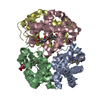
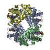
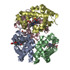
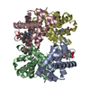
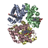
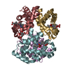
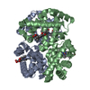
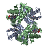
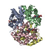
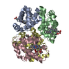
| Title | HORSE METHEMOGLOBIN LOW SALT, PH 7.0 | ||||||
 Components Components |
| ||||||
 Keywords Keywords | OXYGEN STORAGE/TRANSPORT / AQUO METHEMOGLOBIN / QUARTERNARY ASSOCIATION / ALLOSTERIC TRANSITION / OXYGEN STORAGE-TRANSPORT COMPLEX | ||||||
| Function / homology |  Function and homology information Function and homology informationhemoglobin alpha binding / haptoglobin-hemoglobin complex / hemoglobin complex / oxygen transport / erythrocyte development / oxygen carrier activity / oxygen binding / iron ion binding / heme binding / metal ion binding Similarity search - Function | ||||||
| Biological species |  | ||||||
| Method |  X-RAY DIFFRACTION / MOLECULAR REPLACEMENT / Resolution: 3.1 Å X-RAY DIFFRACTION / MOLECULAR REPLACEMENT / Resolution: 3.1 Å | ||||||
 Authors Authors | Sankaranarayanan, R. / Biswal, B.K. / Vijayan, M. | ||||||
 Citation Citation | Journal: Proteins / Year: 2005 Title: A new relaxed state in horse methemoglobin characterized by crystallographic studies. Authors: Sankaranarayanan, R. / Biswal, B.K. / Vijayan, M. #1: Journal: Curr.Sci. / Year: 2001Title: Structure of Human Methaemoglobin: The Variation of a Theme Authors: Biswal, B.K. / Vijayan, M. #2: Journal: Acta Crystallogr.,Sect.D / Year: 2002Title: Structures of Human Oxy- and Deoxyhaemoglobin at Different Levels of Humidity: Variability in the T State Authors: Biswal, B.K. / Vijayan, M. | ||||||
| History |
|
- Structure visualization
| Structure viewer | Molecule: MolmilJmol/JSmol |
|---|
- Downloads & links
-Download
| PDBx/mmCIF format | 1y8h.cif.gz | 118.5 KB | Display | PDBx/mmCIF format |
|---|---|---|---|---|
| PDB format | pdb1y8h.ent.gz | 94.2 KB | Display | PDB format |
| PDBx/mmJSON format | 1y8h.json.gz | Tree view | PDBx/mmJSON format | |
| Others |  Other downloads Other downloads |
-Validation report
| Arichive directory | https://data.pdbj.org/pub/pdb/validation_reports/y8/1y8hftp://data.pdbj.org/pub/pdb/validation_reports/y8/1y8h | HTTPS FTP |
|---|
-Related structure data
| Related structure data | 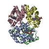 1y8iC 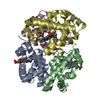 1y8kC 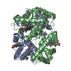 2mhbS C: citing same article ( S: Starting model for refinement |
|---|---|
| Similar structure data |


-Links
 PDBj
PDBj


- Assembly
| Deposited unit | 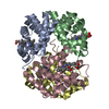
| ||||||||
|---|---|---|---|---|---|---|---|---|---|
| 1 |
| ||||||||
| Unit cell |
|
-Components
| #1: Protein | Mass: 15138.280 Da / Num. of mol.: 2 / Source method: isolated from a natural source / Details: PH 7.0 AQUOMET STRUCTURE / Source: (natural) #2: Protein | Mass: 16032.274 Da / Num. of mol.: 2 / Source method: isolated from a natural source / Source: (natural) #3: Chemical | ChemComp-HEM /   Mass: 616.487 Da / Num. of mol.: 4 / Source method: obtained synthetically / Formula: C34H32FeN4O4 Mass: 616.487 Da / Num. of mol.: 4 / Source method: obtained synthetically / Formula: C34H32FeN4O4#4: Water | ChemComp-HOH / |  Mass: 18.015 Da / Num. of mol.: 136 / Source method: isolated from a natural source / Formula: H2O Mass: 18.015 Da / Num. of mol.: 136 / Source method: isolated from a natural source / Formula: H2O |
|---|
-Experimental details
-Experiment
| Experiment | Method: X-RAY DIFFRACTION / Number of used crystals: 1 |
|---|
- Sample preparation
| Crystal | Density Matthews: 2.3 Å3/Da / Density % sol: 46.1 % |
|---|---|
| Crystal grow | Temperature: 293 K / Method: liquid diffusion / pH: 7 Details: 0.01M Phosphate Buffer, 30%PEG3350, pH 7.0, LIQUID DIFFUSION, temperature 293K |
-Data collection
| Diffraction | Mean temperature: 293 K |
|---|---|
| Diffraction source | Source: ROTATING ANODE / Type: RIGAKU RU200 / Wavelength: 1.5418 / Wavelength: 1.5418 Å |
| Detector | Type: MARRESEARCH / Detector: IMAGE PLATE / Date: Aug 24, 2002 / Details: OSMIC MIRROR |
| Radiation | Monochromator: GRAPHITE / Protocol: SINGLE WAVELENGTH / Monochromatic (M) / Laue (L): M / Scattering type: x-ray |
| Radiation wavelength | Wavelength: 1.5418 Å / Relative weight: 1 |
| Reflection | Resolution: 3.1→20 Å / Num. obs: 10748 / % possible obs: 95.9 % / Observed criterion σ(F): 0 / Observed criterion σ(I): 0 / Biso Wilson estimate: 22 Å2 / Rmerge(I) obs: 0.188 |
| Reflection shell | Resolution: 3.1→3.21 Å / Rmerge(I) obs: 0.511 / Num. unique all: 1028 / % possible all: 93.5 |
- Processing
| Software |
| |||||||||||||||||||||||||
|---|---|---|---|---|---|---|---|---|---|---|---|---|---|---|---|---|---|---|---|---|---|---|---|---|---|---|
| Refinement | Method to determine structure: MOLECULAR REPLACEMENT Starting model: HORSE METHEMOGLOBIN, pdb entry 2MHB Resolution: 3.1→20 Å / Rfactor Rfree error: 0.008 / Data cutoff high absF: 218152.43 / Data cutoff low absF: 0 / Isotropic thermal model: GROUP / Cross valid method: THROUGHOUT / σ(F): 0
| |||||||||||||||||||||||||
| Solvent computation | Solvent model: FLAT MODEL / Bsol: 31.0083 Å2 / ksol: 0.23056 e/Å3 | |||||||||||||||||||||||||
| Displacement parameters | Biso mean: 40.2 Å2
| |||||||||||||||||||||||||
| Refine analyze |
| |||||||||||||||||||||||||
| Refinement step | Cycle: LAST / Resolution: 3.1→20 Å
| |||||||||||||||||||||||||
| Refine LS restraints |
| |||||||||||||||||||||||||
| LS refinement shell | Resolution: 3.1→3.29 Å / Rfactor Rfree error: 0.029 / Total num. of bins used: 6
| |||||||||||||||||||||||||
| Xplor file |
|
