- PDB-1y65: Crystal structure of beta-hexosaminidase from Vibrio cholerae in ... -
+
Open data
ID or keywords:
Loading...
-
Basic information
Entry
Database: PDB / ID: 1y65
Title
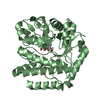
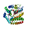
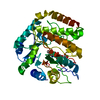
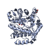
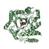
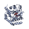
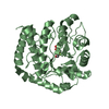
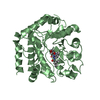
Crystal structure of beta-hexosaminidase from Vibrio cholerae in complex with N-acetyl-D-glucosamine to a resolution of 1.85
Components
Beta-hexosaminidase
Keywords
HYDROLASE / beta-alpha barrel / Structural Genomics / PSI / Protein Structure Initiative / New York SGX Research Center for Structural Genomics / NYSGXRC
Function / homology
Function and homology information
beta-N-acetylhexosaminidase / peptidoglycan turnover / peptidoglycan biosynthetic process / beta-N-acetylglucosaminidase activity / cell wall organization / regulation of cell shape / carbohydrate metabolic process / cell division / response to antibiotic / cytosol Similarity search - Function
Beta-hexosaminidase, bacterial / : / Glycoside hydrolase, family 3, active site / Glycosyl hydrolases family 3 active site. / Glycoside hydrolase, family 3, N-terminal domain / Glycoside hydrolase, family 3, N-terminal / Glycoside hydrolase, family 3, N-terminal domain superfamily / Glycosyl hydrolase family 3 N terminal domain / Glycoside hydrolase superfamily / TIM Barrel ...Beta-hexosaminidase, bacterial / : / Glycoside hydrolase, family 3, active site / Glycosyl hydrolases family 3 active site. / Glycoside hydrolase, family 3, N-terminal domain / Glycoside hydrolase, family 3, N-terminal / Glycoside hydrolase, family 3, N-terminal domain superfamily / Glycosyl hydrolase family 3 N terminal domain / Glycoside hydrolase superfamily / TIM Barrel / Alpha-Beta Barrel / Alpha Beta Similarity search - Domain/homology
Journal: To be Published Title: Crystal structure of beta-hexosaminidase from Vibrio cholerae in complex with N-acetyl-D-glucosamine to a resolution of 1.85 Authors: Gorman, J. / Shapiro, L.
History
Deposition
Dec 3, 2004
Deposition site: RCSB / Processing site: RCSB
Revision 1.0
Dec 14, 2004
Provider: repository / Type: Initial release
Revision 1.1
Apr 30, 2008
Group: Version format compliance
Revision 1.2
Jul 13, 2011
Group: Non-polymer description / Version format compliance
In the structure databanks used in Yorodumi, some data are registered as the other names, "COVID-19 virus" and "2019-nCoV". Here are the details of the virus and the list of structure data.
Jan 31, 2019. EMDB accession codes are about to change! (news from PDBe EMDB page)
EMDB accession codes are about to change! (news from PDBe EMDB page)
The allocation of 4 digits for EMDB accession codes will soon come to an end. Whilst these codes will remain in use, new EMDB accession codes will include an additional digit and will expand incrementally as the available range of codes is exhausted. The current 4-digit format prefixed with “EMD-” (i.e. EMD-XXXX) will advance to a 5-digit format (i.e. EMD-XXXXX), and so on. It is currently estimated that the 4-digit codes will be depleted around Spring 2019, at which point the 5-digit format will come into force.
The EM Navigator/Yorodumi systems omit the EMD- prefix.
Related info.:Q: What is EMD? / ID/Accession-code notation in Yorodumi/EM Navigator
Yorodumi is a browser for structure data from EMDB, PDB, SASBDB, etc.
This page is also the successor to EM Navigator detail page, and also detail information page/front-end page for Omokage search.
The word "yorodu" (or yorozu) is an old Japanese word meaning "ten thousand". "mi" (miru) is to see.
Related info.:EMDB / PDB / SASBDB / Comparison of 3 databanks / Yorodumi Search / Aug 31, 2016. New EM Navigator & Yorodumi / Yorodumi Papers / Jmol/JSmol / Function and homology information / Changes in new EM Navigator and Yorodumi
 Movie
Movie Controller

 Yorodumi
 Open data
 Basic information
 Components
 Keywords
 Function and homology information
 Vibrio cholerae (bacteria)
 X-RAY DIFFRACTION /
  Authors
 Citation
 Structure visualization
 Downloads & links
 Other downloads


 PDBj
PDBj Assembly
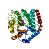

 Type: D-saccharide, beta linking / Mass: 221.208 Da / Num. of mol.: 1
 Mass: 18.015 Da / Num. of mol.: 466 / Source method: isolated from a natural source / Formula: H2O
 Sample preparation
 / Beamline: X4A / Wavelength: 0.9795 Å
 Processing