 Movie
Movie Controller

[English] 日本語
 Yorodumi
- PDB-5utq: Crystal structure of Burkholderia cenocepacia family 3 glycoside ... -
+ Open data

- Basic information
| Entry | Database: PDB / ID: 5utq | ||||||
|---|---|---|---|---|---|---|---|
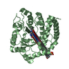
| Title | Crystal structure of Burkholderia cenocepacia family 3 glycoside hydrolase (NagZ) bound to PUGNAc | ||||||
 Components Components | Beta-hexosaminidase | ||||||
 Keywords Keywords | HYDROLASE/HYDROLASE INHIBITOR / Glycoside hydrolase / GH / family 3 / HYDROLASE-HYDROLASE INHIBITOR complex | ||||||
| Function / homology |  Function and homology information Function and homology informationbeta-N-acetylhexosaminidase activity / beta-N-acetylhexosaminidase / peptidoglycan turnover / peptidoglycan biosynthetic process / cell wall organization / regulation of cell shape / carbohydrate metabolic process / cell division / cytoplasm Similarity search - Function | ||||||
| Biological species |  Burkholderia cenocepacia (bacteria) Burkholderia cenocepacia (bacteria) | ||||||
| Method |  X-RAY DIFFRACTION / MOLECULAR REPLACEMENT / Resolution: 2.2 Å X-RAY DIFFRACTION / MOLECULAR REPLACEMENT / Resolution: 2.2 Å | ||||||
 Authors Authors | Vadlamani, G. / Mark, B.L. | ||||||
 Citation Citation | Journal: Protein Sci. / Year: 2017 Title: Conformational flexibility of the glycosidase NagZ allows it to bind structurally diverse inhibitors to suppress beta-lactam antibiotic resistance. Authors: Vadlamani, G. / Stubbs, K.A. / Desire, J. / Bleriot, Y. / Vocadlo, D.J. / Mark, B.L. | ||||||
| History |
|
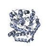
- Structure visualization
| Structure viewer | Molecule: MolmilJmol/JSmol |
|---|
- Downloads & links
-Download
| PDBx/mmCIF format | 5utq.cif.gz | 254 KB | Display | PDBx/mmCIF format |
|---|---|---|---|---|
| PDB format | pdb5utq.ent.gz | 205.3 KB | Display | PDB format |
| PDBx/mmJSON format | 5utq.json.gz | Tree view | PDBx/mmJSON format | |
| Others |  Other downloads Other downloads |
-Validation report
| Arichive directory | https://data.pdbj.org/pub/pdb/validation_reports/ut/5utqftp://data.pdbj.org/pub/pdb/validation_reports/ut/5utq | HTTPS FTP |
|---|
-Related structure data
| Related structure data |  5utpC  5utrC 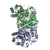 4g6cS S: Starting model for refinement C: citing same article ( |
|---|---|
| Similar structure data |


-Links
 PDBj
PDBj
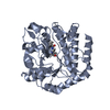
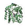
- Assembly
| Deposited unit | 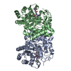
| ||||||||
|---|---|---|---|---|---|---|---|---|---|
| 1 | 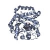
| ||||||||
| 2 | 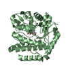
| ||||||||
| Unit cell |
|
-Components
| #1: Protein | Mass: 38116.766 Da / Num. of mol.: 2 Source method: isolated from a genetically manipulated source Source: (gene. exp.) Burkholderia cenocepacia (bacteria) / Gene: nagZ, A3203_21235, WL84_04775 / Production host: References: UniProt: A0A125HFC0, UniProt: B4EA43*PLUS, beta-N-acetylhexosaminidase #2: Chemical |   Mass: 353.327 Da / Num. of mol.: 2 / Source method: obtained synthetically / Formula: C15H19N3O7 Mass: 353.327 Da / Num. of mol.: 2 / Source method: obtained synthetically / Formula: C15H19N3O7#3: Chemical | ChemComp-MES / |   Mass: 195.237 Da / Num. of mol.: 1 / Source method: obtained synthetically / Formula: C6H13NO4S / Comment: pH buffer*YM Mass: 195.237 Da / Num. of mol.: 1 / Source method: obtained synthetically / Formula: C6H13NO4S / Comment: pH buffer*YM#4: Water | ChemComp-HOH / |  Mass: 18.015 Da / Num. of mol.: 359 / Source method: isolated from a natural source / Formula: H2O Mass: 18.015 Da / Num. of mol.: 359 / Source method: isolated from a natural source / Formula: H2O |
|---|
-Experimental details
-Experiment
| Experiment | Method: X-RAY DIFFRACTION / Number of used crystals: 1 |
|---|
- Sample preparation
| Crystal | Density Matthews: 1.87 Å3/Da / Density % sol: 34.08 % |
|---|---|
| Crystal grow | Temperature: 293 K / Method: vapor diffusion, hanging drop / Details: 30-32% PEG8000, 0.1 M MES, pH 6.2-6.8 / PH range: 6.2-6.8 |
-Data collection
| Diffraction | Mean temperature: 100 K |
|---|---|
| Diffraction source | Source: ROTATING ANODE / Type: RIGAKU MICROMAX-007 HF / Wavelength: 1.5418 Å |
| Detector | Type: RIGAKU RAXIS IV++ / Detector: IMAGE PLATE / Date: Sep 13, 2013 |
| Radiation | Protocol: SINGLE WAVELENGTH / Monochromatic (M) / Laue (L): M / Scattering type: x-ray |
| Radiation wavelength | Wavelength: 1.5418 Å / Relative weight: 1 |
| Reflection | Resolution: 2.2→43.83 Å / Num. obs: 28278 / % possible obs: 99.4 % / Redundancy: 2.2 % / CC1/2: 0.966 / Rmerge(I) obs: 0.124 / Net I/σ(I): 6.4 |
| Reflection shell | Resolution: 2.2→2.27 Å / Redundancy: 2.2 % / Rmerge(I) obs: 0.458 / Mean I/σ(I) obs: 2 / Num. unique obs: 2423 / CC1/2: 0.647 / % possible all: 99.2 |
- Processing
| Software |
| ||||||||||||||||||||||||||||||||||||||||||||||||||||||||||||||||||||||||||||||||||||||||||||||||||
|---|---|---|---|---|---|---|---|---|---|---|---|---|---|---|---|---|---|---|---|---|---|---|---|---|---|---|---|---|---|---|---|---|---|---|---|---|---|---|---|---|---|---|---|---|---|---|---|---|---|---|---|---|---|---|---|---|---|---|---|---|---|---|---|---|---|---|---|---|---|---|---|---|---|---|---|---|---|---|---|---|---|---|---|---|---|---|---|---|---|---|---|---|---|---|---|---|---|---|---|
| Refinement | Method to determine structure: MOLECULAR REPLACEMENT Starting model: PDB entry 4G6C Resolution: 2.2→36.302 Å / SU ML: 0.24 / Cross valid method: FREE R-VALUE / σ(F): 1.34 / Phase error: 20.48
| ||||||||||||||||||||||||||||||||||||||||||||||||||||||||||||||||||||||||||||||||||||||||||||||||||
| Solvent computation | Shrinkage radii: 0.9 Å / VDW probe radii: 1.11 Å | ||||||||||||||||||||||||||||||||||||||||||||||||||||||||||||||||||||||||||||||||||||||||||||||||||
| Refinement step | Cycle: LAST / Resolution: 2.2→36.302 Å
| ||||||||||||||||||||||||||||||||||||||||||||||||||||||||||||||||||||||||||||||||||||||||||||||||||
| Refine LS restraints |
| ||||||||||||||||||||||||||||||||||||||||||||||||||||||||||||||||||||||||||||||||||||||||||||||||||
| LS refinement shell |
|
