 Movie
Movie Controller

[English] 日本語
 Yorodumi
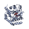
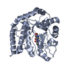
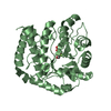
- PDB-3gsm: Vibrio cholerae family 3 glycoside hydrolase (NagZ) bound to N-Va... -
+ Open data

- Basic information
| Entry | Database: PDB / ID: 3gsm | ||||||
|---|---|---|---|---|---|---|---|
| Title | Vibrio cholerae family 3 glycoside hydrolase (NagZ) bound to N-Valeryl-PUGNAc | ||||||
 Components Components | Beta-hexosaminidase | ||||||
 Keywords Keywords | HYDROLASE / glycoside hydrolases / Cell cycle / Cell division / Cell shape / Cell wall biogenesis/degradation / Glycosidase / Peptidoglycan synthesis | ||||||
| Function / homology |  Function and homology information Function and homology informationbeta-N-acetylhexosaminidase / peptidoglycan turnover / peptidoglycan biosynthetic process / beta-N-acetylglucosaminidase activity / cell wall organization / regulation of cell shape / carbohydrate metabolic process / cell division / response to antibiotic / cytosol Similarity search - Function | ||||||
| Biological species |   Vibrio cholerae (bacteria) Vibrio cholerae (bacteria) | ||||||
| Method |  X-RAY DIFFRACTION / MOLECULAR REPLACEMENT / Resolution: 2.4 Å X-RAY DIFFRACTION / MOLECULAR REPLACEMENT / Resolution: 2.4 Å | ||||||
 Authors Authors | Balcewich, M.D. / Mark, B.L. | ||||||
 Citation Citation | Journal: Protein Sci. / Year: 2009 Title: Insight into a strategy for attenuating AmpC-mediated beta-lactam resistance: structural basis for selective inhibition of the glycoside hydrolase NagZ. Authors: Balcewich, M.D. / Stubbs, K.A. / He, Y. / James, T.W. / Davies, G.J. / Vocadlo, D.J. / Mark, B.L. | ||||||
| History |
|
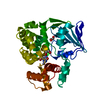
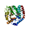
- Structure visualization
| Structure viewer | Molecule: MolmilJmol/JSmol |
|---|
- Downloads & links
-Download
| PDBx/mmCIF format | 3gsm.cif.gz | 80.4 KB | Display | PDBx/mmCIF format |
|---|---|---|---|---|
| PDB format | pdb3gsm.ent.gz | 58.5 KB | Display | PDB format |
| PDBx/mmJSON format | 3gsm.json.gz | Tree view | PDBx/mmJSON format | |
| Others |  Other downloads Other downloads |
-Validation report
| Arichive directory | https://data.pdbj.org/pub/pdb/validation_reports/gs/3gsmftp://data.pdbj.org/pub/pdb/validation_reports/gs/3gsm | HTTPS FTP |
|---|
-Related structure data
| Related structure data |  2wcaC  3gs6C  2oxnS S: Starting model for refinement C: citing same article ( |
|---|---|
| Similar structure data |


-Links
 PDBj
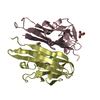
PDBj- Assembly
| Deposited unit | 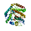
| ||||||||
|---|---|---|---|---|---|---|---|---|---|
| 1 |
| ||||||||
| Unit cell |
| ||||||||
| Details | one biological molecule in the asymmetric unit |
-Components
| #1: Protein | Mass: 37714.848 Da / Num. of mol.: 1 / Mutation: E19A, Q22A, K54A Source method: isolated from a genetically manipulated source Source: (gene. exp.) Vibrio cholerae (bacteria) / Strain: Vibrio cholerae O1 biovar eltor str. N16961 / Gene: family 3 glycoside hydrolase NagZ, nagZ, VC_0692 / Plasmid: pET / Production host: | ||
|---|---|---|---|
| #2: Chemical | ChemComp-VPU / [[(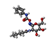  Mass: 395.407 Da / Num. of mol.: 1 / Source method: obtained synthetically / Formula: C18H25N3O7 Mass: 395.407 Da / Num. of mol.: 1 / Source method: obtained synthetically / Formula: C18H25N3O7 | ||
| #3: Chemical |   Mass: 92.094 Da / Num. of mol.: 2 / Source method: obtained synthetically / Formula: C3H8O3 Mass: 92.094 Da / Num. of mol.: 2 / Source method: obtained synthetically / Formula: C3H8O3#4: Water | ChemComp-HOH / |  Mass: 18.015 Da / Num. of mol.: 128 / Source method: isolated from a natural source / Formula: H2O Mass: 18.015 Da / Num. of mol.: 128 / Source method: isolated from a natural source / Formula: H2O |
-Experimental details
-Experiment
| Experiment | Method: X-RAY DIFFRACTION / Number of used crystals: 1 |
|---|
- Sample preparation
| Crystal | Density Matthews: 2.38 Å3/Da / Density % sol: 48.24 % |
|---|---|
| Crystal grow | Temperature: 298 K / Method: vapor diffusion, hanging drop / pH: 6.2 Details: 13% PEG 20,000, 10% glycerol, 100mM Bis-Tris pH 6.2, VAPOR DIFFUSION, HANGING DROP, temperature 298K |
-Data collection
| Diffraction | Mean temperature: 100 K |
|---|---|
| Diffraction source | Source: ROTATING ANODE / Type: RIGAKU RUH2R / Wavelength: 1.54 Å |
| Detector | Type: RIGAKU RAXIS IV++ / Detector: IMAGE PLATE / Date: Oct 20, 2008 |
| Radiation | Protocol: SINGLE WAVELENGTH / Monochromatic (M) / Laue (L): M / Scattering type: x-ray |
| Radiation wavelength | Wavelength: 1.54 Å / Relative weight: 1 |
| Reflection | Resolution: 2.4→61.08 Å / Num. all: 14609 / Num. obs: 14482 / % possible obs: 99.5 % / Observed criterion σ(F): 0 / Observed criterion σ(I): 0 / Redundancy: 2.97 % / Rmerge(I) obs: 0.1 / Net I/σ(I): 6.7 |
| Reflection shell | Resolution: 2.4→2.53 Å / Redundancy: 3 % / Rmerge(I) obs: 0.49 / Mean I/σ(I) obs: 2.2 / % possible all: 100 |
- Processing
| Software |
| ||||||||||||||||||||||||||||||||||||||||||||||||||||||||||||||||||||||||||||||||||||||||||||||||||||||||||||||||||||||||||||||||||||||||||||||||||||||||||||||||||||||||||
|---|---|---|---|---|---|---|---|---|---|---|---|---|---|---|---|---|---|---|---|---|---|---|---|---|---|---|---|---|---|---|---|---|---|---|---|---|---|---|---|---|---|---|---|---|---|---|---|---|---|---|---|---|---|---|---|---|---|---|---|---|---|---|---|---|---|---|---|---|---|---|---|---|---|---|---|---|---|---|---|---|---|---|---|---|---|---|---|---|---|---|---|---|---|---|---|---|---|---|---|---|---|---|---|---|---|---|---|---|---|---|---|---|---|---|---|---|---|---|---|---|---|---|---|---|---|---|---|---|---|---|---|---|---|---|---|---|---|---|---|---|---|---|---|---|---|---|---|---|---|---|---|---|---|---|---|---|---|---|---|---|---|---|---|---|---|---|---|---|---|---|---|
| Refinement | Method to determine structure: MOLECULAR REPLACEMENT Starting model: 2OXN Resolution: 2.4→61.08 Å / Cor.coef. Fo:Fc: 0.925 / Cor.coef. Fo:Fc free: 0.884 / SU B: 9.329 / SU ML: 0.219 / Cross valid method: THROUGHOUT / ESU R: 0.485 / ESU R Free: 0.294 / Stereochemistry target values: MAXIMUM LIKELIHOOD Details: The VPU residue, bound inhibitor N-valeryl PUGNAc, has missing atoms: OAQ CAP OAR NAO CAS CAT CAU CAV CAW CAX. HYDROGENS HAVE BEEN ADDED IN THE RIDING POSITIONS.
| ||||||||||||||||||||||||||||||||||||||||||||||||||||||||||||||||||||||||||||||||||||||||||||||||||||||||||||||||||||||||||||||||||||||||||||||||||||||||||||||||||||||||||
| Solvent computation | Ion probe radii: 0.8 Å / Shrinkage radii: 0.8 Å / VDW probe radii: 1.4 Å / Solvent model: MASK | ||||||||||||||||||||||||||||||||||||||||||||||||||||||||||||||||||||||||||||||||||||||||||||||||||||||||||||||||||||||||||||||||||||||||||||||||||||||||||||||||||||||||||
| Displacement parameters | Biso mean: 26.499 Å2
| ||||||||||||||||||||||||||||||||||||||||||||||||||||||||||||||||||||||||||||||||||||||||||||||||||||||||||||||||||||||||||||||||||||||||||||||||||||||||||||||||||||||||||
| Refinement step | Cycle: LAST / Resolution: 2.4→61.08 Å
| ||||||||||||||||||||||||||||||||||||||||||||||||||||||||||||||||||||||||||||||||||||||||||||||||||||||||||||||||||||||||||||||||||||||||||||||||||||||||||||||||||||||||||
| Refine LS restraints |
| ||||||||||||||||||||||||||||||||||||||||||||||||||||||||||||||||||||||||||||||||||||||||||||||||||||||||||||||||||||||||||||||||||||||||||||||||||||||||||||||||||||||||||
| LS refinement shell | Resolution: 2.4→2.462 Å / Total num. of bins used: 20
|
