 Movie
Movie Controller


+ Open data

- Basic information
| Entry | Database: PDB / ID: 1y44 | ||||||
|---|---|---|---|---|---|---|---|
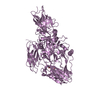
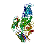
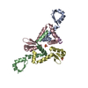
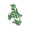
| Title | Crystal structure of RNase Z | ||||||
 Components Components | Ribonuclease Z | ||||||
 Keywords Keywords | HYDROLASE / ZINC-DEPENDENT METAL HYDROLASE | ||||||
| Function / homology |  Function and homology information Function and homology informationtRNase Z / 3'-tRNA processing endoribonuclease activity / zinc ion binding / identical protein binding Similarity search - Function | ||||||
| Biological species |  | ||||||
| Method |  X-RAY DIFFRACTION / SYNCHROTRON / MAD / Resolution: 2.1 Å X-RAY DIFFRACTION / SYNCHROTRON / MAD / Resolution: 2.1 Å | ||||||
 Authors Authors | de la Sierra-Gallay, I.L. / Pellegrini, O. / Condon, C. | ||||||
 Citation Citation | Journal: Nature / Year: 2005 Title: Structural basis for substrate binding, cleavage and allostery in the tRNA maturase RNase Z. Authors: de la Sierra-Gallay, I.L. / Pellegrini, O. / Condon, C. #1: Journal: Embo J. / Year: 2003Title: Endonucleolytic processing of CCA-less tRNA precursors by RNase Z in Bacillus subtilis Authors: Pellegrini, O. / Nezzar, J. / Marchfelder, A. / Putzer, H. / Condon, C. | ||||||
| History |
|
- Structure visualization
| Structure viewer | Molecule: MolmilJmol/JSmol |
|---|
- Downloads & links
-Download
| PDBx/mmCIF format | 1y44.cif.gz | 128.4 KB | Display | PDBx/mmCIF format |
|---|---|---|---|---|
| PDB format | pdb1y44.ent.gz | 99 KB | Display | PDB format |
| PDBx/mmJSON format | 1y44.json.gz | Tree view | PDBx/mmJSON format | |
| Others |  Other downloads Other downloads |
-Validation report
| Arichive directory | https://data.pdbj.org/pub/pdb/validation_reports/y4/1y44ftp://data.pdbj.org/pub/pdb/validation_reports/y4/1y44 | HTTPS FTP |
|---|
-Related structure data
| Similar structure data |
|---|


-Links
 PDBj
PDBj

- Assembly
| Deposited unit | 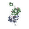
| ||||||||
|---|---|---|---|---|---|---|---|---|---|
| 1 |
| ||||||||
| Unit cell |
|
-Components
-Protein , 1 types, 2 molecules AB
| #1: Protein | Mass: 35867.066 Da / Num. of mol.: 2 / Mutation: I46M, L228M Source method: isolated from a genetically manipulated source Source: (gene. exp.) |
|---|
-Non-polymers , 5 types, 183 molecules 


| #2: Chemical |  Mass: 65.409 Da / Num. of mol.: 2 / Source method: obtained synthetically / Formula: Zn Mass: 65.409 Da / Num. of mol.: 2 / Source method: obtained synthetically / Formula: Zn#3: Chemical | ChemComp-PO4 / |  Mass: 94.971 Da / Num. of mol.: 1 / Source method: obtained synthetically / Formula: PO4 Mass: 94.971 Da / Num. of mol.: 1 / Source method: obtained synthetically / Formula: PO4#4: Chemical |  Mass: 92.094 Da / Num. of mol.: 3 / Source method: obtained synthetically / Formula: C3H8O3 Mass: 92.094 Da / Num. of mol.: 3 / Source method: obtained synthetically / Formula: C3H8O3#5: Chemical | ChemComp-MES / |  Mass: 195.237 Da / Num. of mol.: 1 / Source method: obtained synthetically / Formula: C6H13NO4S / Comment: pH buffer*YM Mass: 195.237 Da / Num. of mol.: 1 / Source method: obtained synthetically / Formula: C6H13NO4S / Comment: pH buffer*YM#6: Water | ChemComp-HOH / | Mass: 18.015 Da / Num. of mol.: 176 / Source method: isolated from a natural source / Formula: H2O |
|---|
-Details
| Has protein modification | Y |
|---|
-Experimental details
-Experiment
| Experiment | Method: X-RAY DIFFRACTION / Number of used crystals: 1 |
|---|
- Sample preparation
| Crystal | Density Matthews: 2.6 Å3/Da / Density % sol: 52 % |
|---|---|
| Crystal grow | Temperature: 293 K / Method: vapor diffusion, hanging drop / pH: 6 Details: PEG MME 2000, ammonium sulfate, sodium-MES, VAPOR DIFFUSION, HANGING DROP, temperature 293K, pH 6 |
-Data collection
| Diffraction | Mean temperature: 100 K | ||||||||||||
|---|---|---|---|---|---|---|---|---|---|---|---|---|---|
| Diffraction source | Source: SYNCHROTRON / Site: ESRF  / Beamline: BM30A / Wavelength: 0.9766, 0.9796, 0.9797 / Beamline: BM30A / Wavelength: 0.9766, 0.9796, 0.9797 | ||||||||||||
| Detector | Type: MARRESEARCH / Detector: CCD / Date: Apr 29, 2004 / Details: mirrors | ||||||||||||
| Radiation | Monochromator: 2 SILICON CRYSTALS / Protocol: MAD / Monochromatic (M) / Laue (L): M / Scattering type: x-ray | ||||||||||||
| Radiation wavelength |
| ||||||||||||
| Reflection | Resolution: 2.05→50 Å / Num. obs: 85742 / Observed criterion σ(I): -3 / Redundancy: 2.32 % / Biso Wilson estimate: 21.5 Å2 / Rmerge(I) obs: 0.061 / Net I/σ(I): 8 | ||||||||||||
| Reflection shell | Resolution: 2.05→2.1 Å / Redundancy: 2.1 % / Rmerge(I) obs: 0.415 / Mean I/σ(I) obs: 0.49 / % possible all: 79.3 |
- Processing
| Software |
| ||||||||||||||||||||||||||||||||||||||||||||||||||||||||||||||||||||||||||||||||
|---|---|---|---|---|---|---|---|---|---|---|---|---|---|---|---|---|---|---|---|---|---|---|---|---|---|---|---|---|---|---|---|---|---|---|---|---|---|---|---|---|---|---|---|---|---|---|---|---|---|---|---|---|---|---|---|---|---|---|---|---|---|---|---|---|---|---|---|---|---|---|---|---|---|---|---|---|---|---|---|---|---|
| Refinement | Method to determine structure: MAD / Resolution: 2.1→34.78 Å / Rfactor Rfree error: 0.003 / Occupancy max: 1 / Occupancy min: 1 / Data cutoff high absF: 2976258.97 / Data cutoff low absF: 0 / Isotropic thermal model: RESTRAINED / Cross valid method: THROUGHOUT / σ(F): 0
| ||||||||||||||||||||||||||||||||||||||||||||||||||||||||||||||||||||||||||||||||
| Solvent computation | Solvent model: FLAT MODEL / Bsol: 53.5034 Å2 / ksol: 0.380324 e/Å3 | ||||||||||||||||||||||||||||||||||||||||||||||||||||||||||||||||||||||||||||||||
| Displacement parameters | Biso mean: 37.6 Å2
| ||||||||||||||||||||||||||||||||||||||||||||||||||||||||||||||||||||||||||||||||
| Refine analyze |
| ||||||||||||||||||||||||||||||||||||||||||||||||||||||||||||||||||||||||||||||||
| Refinement step | Cycle: LAST / Resolution: 2.1→34.78 Å
| ||||||||||||||||||||||||||||||||||||||||||||||||||||||||||||||||||||||||||||||||
| Refine LS restraints |
| ||||||||||||||||||||||||||||||||||||||||||||||||||||||||||||||||||||||||||||||||
| LS refinement shell | Resolution: 2.1→2.23 Å / Rfactor Rfree error: 0.01 / Total num. of bins used: 6
| ||||||||||||||||||||||||||||||||||||||||||||||||||||||||||||||||||||||||||||||||
| Xplor file |
|
